. 2012 Dec;86(24):13324-33.
doi: 10.1128/JVI.01757-12. Epub 2012 Sep 26.

Differential activation of cellular DNA damage responses by replication-defective and replication-competent adenovirus mutants

Affiliations

Differential activation of cellular DNA damage responses by replication-defective and replication-competent adenovirus mutants

Anand Prakash et al. J Virol. 2012 Dec.

Abstract

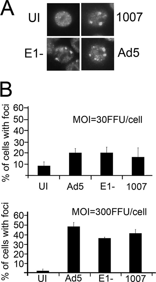
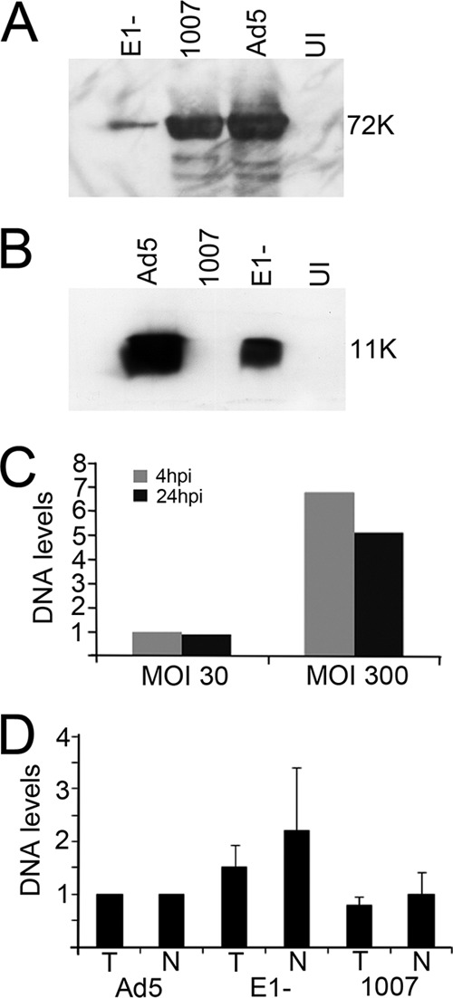
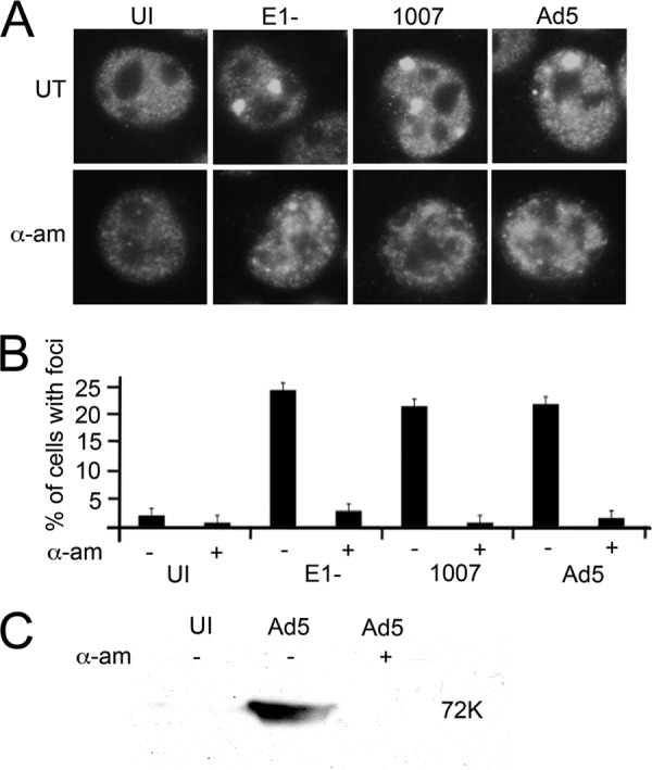
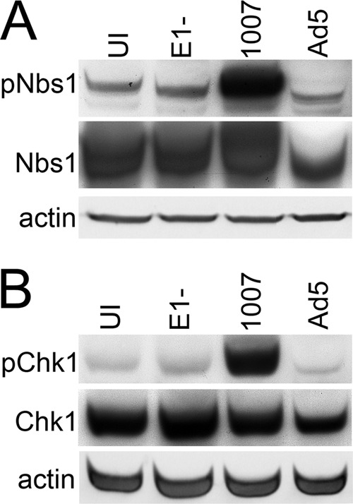
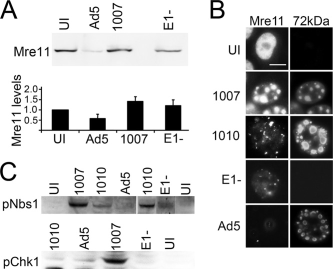
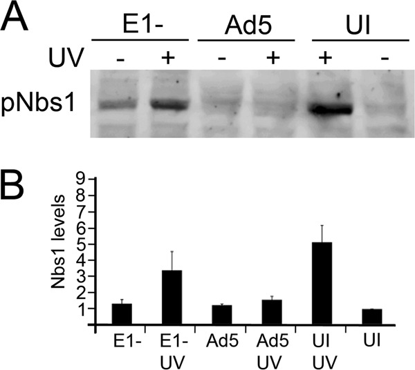
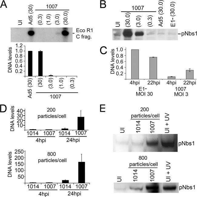
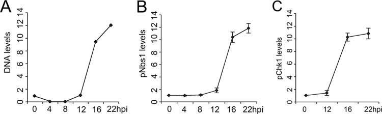
Adenovirus (Ad) mutants that lack early region 4 (E4) activate the phosphorylation of cellular DNA damage response proteins. In wild-type Ad type 5 (Ad5) infections, E1b and E4 proteins target the cellular DNA repair protein Mre11 for redistribution and degradation, thereby interfering with its ability to activate phosphorylation cascades important during DNA repair. The characteristics of Ad infection that activate cellular DNA repair processes are not yet well understood. We investigated the activation of DNA damage responses by a replication-defective Ad vector (AdRSVβgal) that lacks E1 and fails to produce the immediate-early E1a protein. E1a is important for activating early gene expression from the other viral early transcription units, including E4. AdRSVβgal can deliver its genome to the cell, but it is subsequently deficient for viral early gene expression and DNA replication. We studied the ability of AdRSVβgal-infected cells to induce cellular DNA damage responses. AdRSVβgal infection does activate formation of foci containing the Mdc1 protein. However, AdRSVβgal fails to activate phosphorylation of the damage response proteins Nbs1 and Chk1. We found that viral DNA replication is important for Nbs1 phosphorylation, suggesting that this step in the viral life cycle may provide an important trigger for activating at least some DNA repair proteins.


Figures

Fig 1
Fig 1
Mdc1 focus formation in Ad-infected cells is correlated with MOI. HeLa cells were either uninfected (UI) or infected with Ad5, H5dl1007, or AdRSVβgal (E1−) at an MOI of 30 or 300 FFU/cell as indicated. The cells were then fixed with paraformaldehyde at 5 hpi and immunostained with antibody against Mdc1. (A) Immunofluorescence micrographs showing Mdc1 redistribution into large foci in cells infected with the indicated viruses at an MOI of 30 FFU/cell. (B) Cell cultures were infected with the indicated viruses, blindly scored for Mdc1 foci, and the percentages of cells with large Mdc1 foci were plotted for infections performed at an MOI of 30 or 300 FFU/cell. Error bars show the standard errors of the mean from three independent experiments.
Fig 2
Fig 2
AdRSVβgal is defective for viral early gene expression and viral DNA replication. HeLa cells were either uninfected (UI) or infected with Ad5, H5dl1007, or AdRSVβgal (E1−) at an MOI of 30 FFU/cell. (A and B) Western blotting was performed on extracts prepared at 24 hpi with antibody against the E2-72kDa (72K) (A) and E4-11kDa (11K) (B). (C) Total DNA samples from cells infected with AdRSVβgal at an MOI of 30 or 300 FFU/cell were prepared at 4 and 24 hpi and analyzed by dot blotting. DNA levels were expressed as the fold difference from AdRSVβgal DNA levels measured at 4 hpi for an MOI of 30 FFU/cell, which was set at 1. (D) HeLa cells were infected with the indicated viruses at an MOI of 30 FFU/cell. Total (T) and nuclear (N) DNA samples were prepared at 4 hpi (prior to the onset of DNA replication) and analyzed by dot blotting. DNA levels were expressed relative to the Ad5 levels for both total and nuclear DNA, which were each set at 1. Error bars show the standard errors of the mean from three independent experiments.
Fig 3
Fig 3
Mdc1 focus formation depends on transcription from the viral genome. (A) HeLa cells were either uninfected (UI) or infected with Ad5, H5dl1007, or AdRSVβgal (E1−) viruses at an MOI of 30 FFU/cell. At 30 min postinfection, experimental cells were left untreated or treated with 40 μg of α-amanitin/ml (α-am). The cells were fixed with paraformaldehyde at 5 hpi and immunostained with antibody against Mdc1. (B) Cells in each infected culture were blindly scored for Mdc1 focus formation in three independent experiments, and the results are graphed. Error bars show the standard errors of the mean. (C) As a control, we measured the effect of α-amanitin treatment on Ad5 early gene expression. Ad5-infected HeLa cells were left untreated or treated with α-amanitin from 30 min postinfection. Protein extracts prepared at 10 hpi were subjected to Western blot analysis with an antibody that detect E2-72kDa (72K) as a marker for early gene expression.
Fig 4
Fig 4
Replication-defective AdRSVβgal fails to activate phosphorylation of Nbs1 and Chk1. HeLa cells were either uninfected (UI) or infected with Ad5, H5dl1007, or AdRSVβgal (E1−) at an MOI of 30 FFU/cell for 22 to 24 h. Protein extracts prepared from these cultures were subjected to SDS-PAGE and Western blot analyses using antibodies against phosphorylated Nbs1 (pNbs1) (A) or phosphorylated Chk1 (pChk1) (B). Extracts from the same infections were assayed in Western blots with antibodies to detect unphosphorylated epitopes of Nbs1 and Chk1, and actin (middle and lower panels, respectively).
Fig 5
Fig 5
Redistribution of Mre11 to nuclear tracks is not sufficient to prevent Nbs1 phosphorylation. HeLa cells were either uninfected (UI) or infected with Ad5, H5dl1007, H5dl1010, or AdRSVβgal (E1−) viruses at an MOI of 30 FFU/cell. (A) Western blotting was performed on extracts prepared at 24 hpi from the indicated infections, using antibody against Mre11 (top panel). Phosphorimaging analysis of three independent blots was performed, and the results were plotted (bottom panel). Error bars show the standard error of the mean. (B) Mre11 localization was determined by double immunofluorescence staining of cells infected with the indicated viruses and fixed at 20 to 24 hpi and immunostained with Mre11 and E2-72kDa antibodies. (C) Western blotting was performed on extracts prepared at 24 hpi from the indicated infections, using antibody against phosphorylated Nbs1 (pNbs1) (top panel) or phosphorylated Chk1 (pChk1) (bottom panel).
Fig 6
Fig 6
UV treatment of AdRSVβgal-infected cells activates the phosphorylation of Nbs1. HeLa cells were either left uninfected (UI) or infected with Ad5 or AdRSVβgal (E1−) at an MOI of 30 FFU/cell, and after 22 h the cultures were exposed to UV (100 mJ/cm2) as indicated or left untreated. Cultures were harvested at 24 hpi, and protein lysates were prepared and analyzed by Western blotting with antibody against phosphorylated Nbs1 (pNbs1). A representative Western blot is shown in panel A. Phosphorimaging analysis of three independent blots was performed, and a graph of the results is shown in panel B. Error bars show the standard errors of the mean.
Fig 7
Fig 7
Viral DNA replication is important for activating Nbs1 phosphorylation. HeLa cells were either uninfected (UI) or infected with wild-type Ad5, AdRSVβgal (E1-), H5dl1007, or H5dl1014 at the indicated MOIs and cultured for 22 to 24 hpi. The MOIs used are in parentheses in FFU/cell. (A) Southern blot analysis was performed with 10 μg of EcoRI-digested DNA prepared from each infection. The EcoRI C fragment used for comparison of viral DNA levels is shown in the top panel. Viral DNA levels were quantified by phosphorimaging analysis (lower graph). E4 mutant H5dl1007 DNA levels are expressed as the fraction of the Ad5 level, which was set at 1. Error bars indicate the standard deviations from three independent experiments. (B) Protein extracts from infected cells were subjected to SDS-PAGE and Western blot analysis with antibody against phosphorylated Nbs1. (C) Cells were infected with AdRSVβgal (E1−) and H5dl1007 at MOIs of 30 and 3 FFU/cell, respectively. DNA levels from the indicated infections and times (hpi) were measured by dot blot analysis and are expressed as the fraction of AdRSVβgal DNA levels measured at 4 hpi, which was set at 1. Error bars indicate the standard deviations from three independent experiments. (D) Total DNA samples were prepared from the indicated infections and hpi, and the samples were analyzed by dot blotting. DNA levels were expressed as the fold difference from H5dl1007 DNA levels measured at 4 hpi after infection with 200 particles/cell (top graph) or 800 particles/cell (bottom graph), which was set at 1. Error bars show the standard errors of the mean from three independent experiments. (E) Protein extracts from the indicated infections were subjected to SDS-PAGE and Western blot analysis with antibody against phosphorylated Nbs1 (pNbs1). In panels D and E, MOIs were expressed as virion particles/cell (see Materials and Methods). UV-treated uninfected cells were used as a positive control for induction of pNbs1.
Fig 8
Fig 8
E4 mutant-induced phosphorylation of Nbs1 and Chk1 correlates with the onset of viral DNA replication. HeLa cells were infected with H5dl1007 at 30 FFU/cell. Total DNA was isolated at 0, 4, 8, 16, and 22 hpi for Southern blot analysis and DNA levels were quantified by phosphorimaging analysis. (A) E4 mutant DNA levels were measured as the fold increase over the background level detected in samples prepared from uninfected cells, which was set at 1. In a parallel experiment, protein lysates were prepared from H5dl1007-infected cells at the times indicated and processed for Western blot analyses with antibodies against phosphorylated Nbs1 (B) and Chk1 (C). Protein levels were quantified by phosphorimaging analysis. The levels of phosphorylated Nbs1 (pNbs1) and Chk1 (pChk1) are expressed as the fold increase compared to the level detected in uninfected HeLa cells, which was set at 1.

Similar articles

Cited by

References

    1. Araujo FD, Stracker TH, Carson CT, Lee DV, Weitzman MD. 2005. Adenovirus type 5 E4orf3 protein targets the Mre11 complex to cytoplasmic aggresomes. J. Virol. 79:11382–11391 - PMC - PubMed
    1. Assenmacher N, Hopfner KP. 2004. MRE11/RAD50/NBS1: complex activities. Chromosoma 113:157–166 - PubMed
    1. Baker A, Rohleder KJ, Hanakahi L, Ketner G. 2007. Adenovirus E4 34k and E1b 55k oncoproteins target host DNA ligase IV for proteasomal degradation. J. Virol. 81:7034–7040 - PMC - PubMed
    1. Bakkenist CJ, Kastan MB. 2003. DNA damage activates ATM through intermolecular autophosphorylation and dimer dissociation. Nature 421:499–506 - PubMed
    1. Berk AJ, Lee F, Harrison T, Williams J, Sharp PA. 1979. Pre-early adenovirus 5 gene product regulates synthesis of early viral messenger RNAs. Cell 17:935–944 - PubMed

Publication types