. 2012 Dec;86(24):13445-55.
doi: 10.1128/JVI.01682-12. Epub 2012 Sep 26.

Interferon-inducible protein Mx1 inhibits influenza virus by interfering with functional viral ribonucleoprotein complex assembly

Affiliations

Interferon-inducible protein Mx1 inhibits influenza virus by interfering with functional viral ribonucleoprotein complex assembly

Judith Verhelst et al. J Virol. 2012 Dec.

Abstract

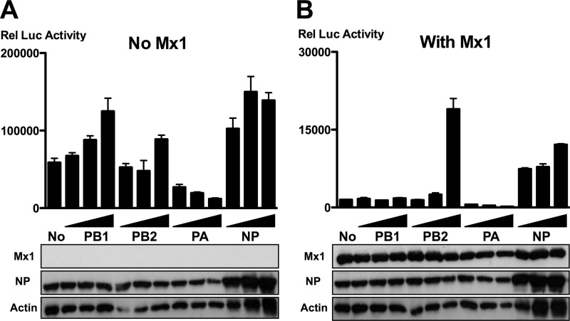
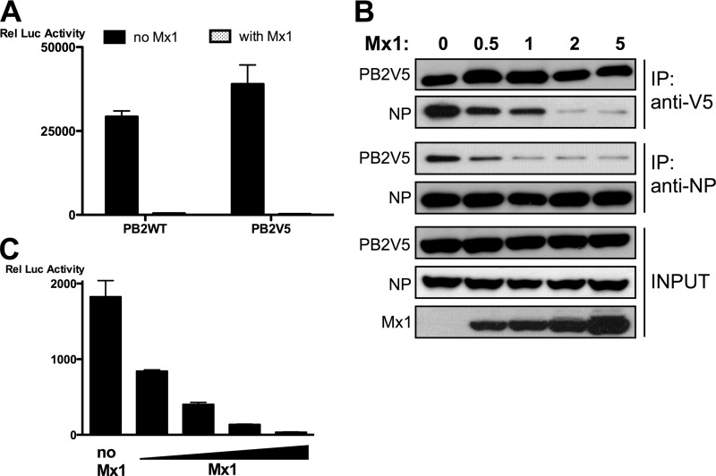
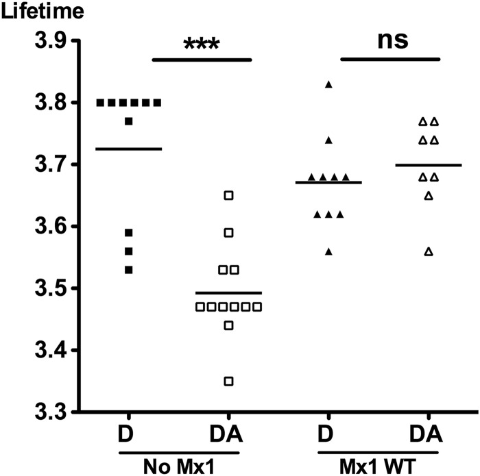
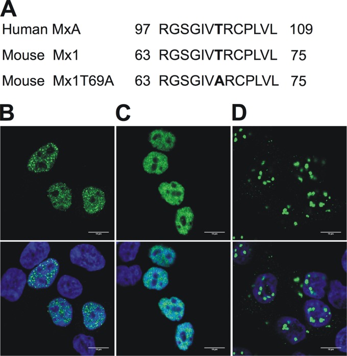
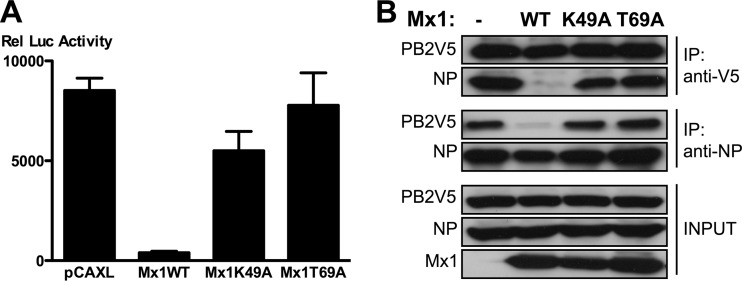
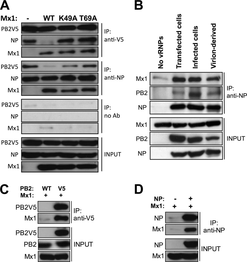
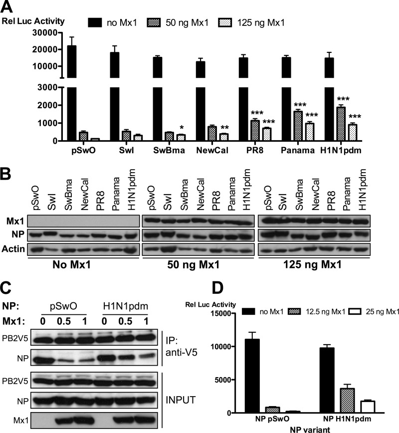
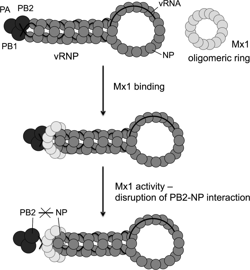
Mx1 is a GTPase that is part of the antiviral response induced by type I and type III interferons in the infected host. It inhibits influenza virus infection by blocking viral transcription and replication, but the molecular mechanism is not known. Polymerase basic protein 2 (PB2) and nucleoprotein (NP) were suggested to be the possible target of Mx1, but a direct interaction between Mx1 and any of the viral proteins has not been reported. We investigated the interplay between Mx1, NP, and PB2 to identify the mechanism of Mx1's antiviral activity. We found that Mx1 inhibits the PB2-NP interaction, and the strength of this inhibition correlated with a decrease in viral polymerase activity. Inhibition of the PB2-NP interaction is an active process requiring enzymatically active Mx1. We also demonstrate that Mx1 interacts with the viral proteins NP and PB2, which indicates that Mx1 protein has a direct effect on the viral ribonucleoprotein complex. In a minireplicon system, avian-like NP from swine virus isolates was more sensitive to inhibition by murine Mx1 than NP from human influenza A virus isolates. Likewise, murine Mx1 displaced avian NP from the viral ribonucleoprotein complex more easily than human NP. The stronger resistance of the A/H1N1 pandemic 2009 virus against Mx1 also correlated with reduced inhibition of the PB2-NP interaction. Our findings support a model in which Mx1 interacts with the influenza ribonucleoprotein complex and interferes with its assembly by disturbing the PB2-NP interaction.


Figures

Fig 1
Fig 1
PB2 and NP outcompete Mx1 activity. HEK293T cells were transfected in triplicate with PB1, PB2, PA, and NP expression plasmids (25 ng each), together with pHW-NSLuc (100 ng) and pRL-CMV (25 ng). In addition, 125 ng of pCAXL (A) or pCAXL-Mx1 (B) was cotransfected. Increasing amounts of PB1, PB2, PA, or NP were introduced by increasing the amount of the corresponding expression vectors (125 ng, 250 ng, or 625 ng). The normalized luciferase activity in the lysates was determined 48 h after transfection. Bars represent the average of the triplicates, and the error bars depict one standard deviation. This graph is representative of three independent experiments. Mx1, NP, and actin expression were determined in the lysates by Western blotting.
Fig 2
Fig 2
Mx1 inhibits the interaction between PB2 and NP. (A) HEK293T cells were transfected in triplicate with PB1, PA, and NP expression plasmids (25 ng each), together with pHW-NSLuc (100 ng) and pRL-CMV (25 ng). In addition, 25 ng pCAXL-PB2 or pCAXL-PB2V5 and 125 ng of pCAXL or pCAXL-Mx1 were cotransfected. The relative luciferase activity in the lysates was determined 48 h after transfection. Bars represent the average of the triplicates, and the error bars depict one standard deviation. This graph is representative of two independent experiments. (B) HEK293T cells were transfected with plasmids for expression of PB1, PB2V5, PA, and NP (1 μg each) and with pHW-NSLuc (1 μg). In addition, increasing amounts of pCAXL-Mx1 were cotransfected (0 μg, 0.5 μg, 1 μg, 2 μg, or 5 μg). Total lysates were made 24 h after transfection, and PB2V5 and NP were immunoprecipitated (IP) with anti-V5 and anti-NP antibodies, respectively. Proteins were visualized by Western blotting with antibodies recognizing the V5 tag, NP (anti-RNP antibody), and Mx1. (C) HEK293T cells were transfected in triplicate with plasmids for expression of PB1, PB2, PA, and NP (25 ng each), together with pHW-NSLuc (100 ng) and pRL-CMV (25 ng). Increasing amounts of pCAXL-Mx1 were cotransfected (0 ng, 12.5 ng, 25 ng, 50 ng, or 125 ng). The normalized luciferase activity in the lysates was determined 48 h after transfection. Bars represent the average of triplicates, and the error bars depict one standard deviation.
Fig 3
Fig 3
Mx1 inhibits the interaction between PB2 and NP as determined by FLIM. HEK293T cells were transfected with 25 ng each of plasmids for expression of PB1, PB2V5, PA, and NP and pHW-NSLuc. In addition, 125 ng of pCAXL or pCAXL-Mx1 was cotransfected. After 24 h, cells were fixed and stained for PB2V5 (anti-V5 antibody with Alexa Fluor 488 as FLIM donor) and NP (anti-NP antibody with Alexa Fluor 555 as FLIM acceptor). For each setting, FLIM measurements were performed on 8 to 12 fields (altogether about 30 cells) and the mean fluorescence lifetime per field was determined. For each setting, the average of the means was calculated and statistical analysis was performed with a Kruskal-Wallis test (***, P < 0.001). D, donor only; DA, donor and acceptor.
Fig 4
Fig 4
GTPase-deficient Mx1 mutants. (A) Alignment of the amino acid sequence of the human MxAT103A mutant, wild-type mouse Mx1, and the mouse Mx1T69A mutant. The mutation is localized between the first and second GTP-binding consensus motifs. Numbers denote the position in the primary amino acid sequence. (B to D) Subcellular localization of Mx1 (B), Mx1K49A (C), and Mx1T69A (D). HEK293T cells were transfected with pCAXL-Mx1WT, -Mx1K49A, or -Mx1T69A (50 ng). After 24 h, cells were fixed and stained with Hoechst stain (DNA, blue, not shown) and anti-Mx1 antibody (green, top). An overlay was made of the two images (bottom). Scale bar, 10 μm.
Fig 5
Fig 5
An intact GTPase domain of Mx1 is required to inhibit the PB2-NP interaction. (A) HEK293T cells were transfected in triplicate with PB1, PB2, PA, and NP expression plasmids (25 ng each), pHW-NSLuc (100 ng), and pRL-CMV (25 ng). Empty control and expression vectors for the different Mx1 mutants were cotransfected (50 ng of pCAXL, 50 ng of pCAXL-Mx1WT, 100 ng of pCAXL-Mx1K49A, or 50 ng of pCAXL-Mx1T69A). After 48 h, the relative luciferase activity in the lysates was determined. Bars represent the average of triplicates, and the error bars depict one standard deviation. This graph is representative of at least three independent experiments. (B) HEK293T cells were transfected with PB1, PB2V5, PA, and NP expression plasmids (0.5 μg each) and with pHW-NSLuc (0.5 μg). Empty control and expression vectors for the different Mx1 mutants were cotransfected (2 μg of pCAXL, 1 μg of pCAXL-Mx1WT, 2 μg of pCAXL-Mx1K49A, or 1 μg of pCAXL-Mx1T69A). Total lysates were made 24 h after transfection, and PB2V5 and NP were immunoprecipitated with anti-V5 and anti-NP antibodies, respectively. Proteins were visualized by Western blotting with antibodies directed against the V5 tag, NP (anti-RNP antibody), or Mx1.
Fig 6
Fig 6
Mx1 interacts with PB2 and NP. (A) HEK293T cells were transfected with PB1, PB2V5, PA, and NP expression plasmids (0.5 μg each) and with pHW-NSLuc (0.5 μg). Empty control and expression vectors for the different Mx1 mutants were cotransfected (2 μg of pCAXL, 1 μg of pCAXL-Mx1WT, 2 μg of pCAXL-Mx1K49A, or 1 μg of pCAXL-Mx1T69A). After 24 h, total lysates were made in the presence of 25 mM N-ethylmaleimide, and PB2V5 and NP were immunoprecipitated with anti-V5 and anti-NP antibodies, respectively. We also included a control immunoprecipitation in the absence of monoclonal antibody (no Ab). Proteins were visualized by Western blotting with antibodies recognizing the V5 tag, NP (anti-RNP antibody), and Mx1. The results shown are representative of two independent experiments. (B) HEK293T cells were transfected with 2.5 μg pCAXL-Mx1 (Mx1 lysates), with 2.5 μg pCAXL (lysates without vRNPs), or with 1 μg of pCAXL-PB1, -PB2V5, -PA, and -NP and 1 μg pHW-NSLuc (lysates with transfected vRNPs). To obtain lysates containing vRNPs from infected cells, HEK293T cells were infected for 4 h with A/PR/8/34 (MOI of 10). vRNPs isolated from virions were generated by lysing influenza A virions (A/PR/8/34, 6.25 × 10⁸ PFU/ml lysis buffer). Lysates from all conditions were made in the presence of 25 mM N-ethylmaleimide. The mixed lysates (Mx1 plus vRNPs) were immunoprecipitated with monoclonal anti-NP antibody. Proteins were visualized by Western blotting with antibodies recognizing NP (anti-RNP antibody), PB2, or Mx1. The results shown are representative of three independent experiments. (C) HEK293T cells were transfected with 2 μg pCAXL-Mx1 and 1 μg pCAXL-PB2V5 or -PB2WT as control. After 24 h, total lysates were made in the presence of 25 mM N-ethylmaleimide, and PB2V5 was immunoprecipitated with anti-V5 antibody. Proteins were visualized by Western blotting with antibodies recognizing the V5 tag, PB2, and Mx1. The results shown are representative of two independent experiments. (D) HEK293T cells were transfected with 2 μg pCAXL-Mx1 and 1 μg pCAXL-NP or pCAXL as control. After 24 h, total lysates were made in the presence of 25 mM N-ethylmaleimide, and NP was immunoprecipitated with anti-NP antibody. Proteins were visualized by Western blotting with antibodies recognizing NP (anti-RNP antibody) and Mx1. The results shown are representative of two independent experiments.
Fig 7
Fig 7
Natural NP variants display differential sensitivity to Mx1. (A and B) HEK293T cells were transfected in triplicate with expression plasmids for PB1, PB2, and PA (25 ng each), pHW-NSLuc (100 ng), and pRL-CMV (25 ng). In addition, 25 ng of expression plasmids for different NP variants were cotransfected (pSwO, A/Swine/Ontario/42729A/01; SwI, A/Swine/Iowa/4/1976; SwBma, mouse-adapted A/Swine/Belgium/1/98; NewCal, A/New Caledonia/20/99; PR8, A/Puerto Rico/8/34; Panama, A/Panama/2007/99; H1N1pdm, A/Mexico/InDRE4487/2009). Increasing amounts of pCAXL-Mx1 were cotransfected (0 ng, 50 ng, and 125 ng). (A) The normalized luciferase activity in the lysates was determined 48 h after transfection. Bars represent the average of triplicates, and the error bars depict one standard deviation. This graph is representative of three independent experiments. In the absence of Mx1 (black bars), no significant difference between the NP variants was observed (P = 0.0528). In the presence of Mx1, the NP variants differed significantly in their sensitivity to the inhibitory activity of Mx1. Results for all groups were compared with results for the most sensitive variant, NP-pSwO. NP variants whose results differ significantly are indicated (*, P < 0.05; **, P < 0.01; ***, P < 0.001). Statistical analysis was performed with a nonparametric one-way analysis of variance followed by Bonferroni's multiple comparison test. (B) Mx1, NP (polyclonal anti-RNP antibody), and actin expression were determined in the lysates by Western blotting. (C) HEK293T cells were transfected with 1 μg each of expression plasmids for PB1, PB2V5, PA, and NP (from pSwO or H1N1pdm) and with 1 μg pHW-NSLuc. Increasing amounts of pCAXL-Mx1 were cotransfected (0 μg, 0.5 μg, and 1 μg). Total lysates were made 24 h later, and PB2V5 was immunoprecipitated by an anti-V5-HRP antibody. Proteins were visualized by Western blotting with antibodies specific for the V5 tag, NP (anti-RNP antibody), and Mx1. (D) HEK293T cells were transfected in triplicate with expression plasmids for PB1, PB2, and PA (25 ng each), together with pHW-NSLuc (100 ng) and pRL-CMV (25 ng). In addition, 25 ng of pCAXL-NP-pSwO or 25 ng of pCAXL-NP-H1N1pdm was cotransfected. Increasing amounts of pCAXL-Mx1 were cotransfected (0 ng, 12.5 ng, and 25 ng). The normalized luciferase activity in the lysates was determined 48 h after transfection. Bars represent the average of triplicates, and the error bars depict one standard deviation.
Fig 8
Fig 8
Proposed mechanism of Mx1's antiviral activity against influenza A virus.

References

    1. Biswas SK, Boutz PL, Nayak DP. 1998. Influenza virus nucleoprotein interacts with influenza virus polymerase proteins. J. Virol. 72:5493–5501 - PMC - PubMed
    1. Boivin S, Hart DJ. 2011. Interaction of the influenza A virus polymerase PB2 C-terminal region with importin alpha isoforms provides insights into host adaptation and polymerase assembly. J. Biol. Chem. 286:10439–10448 - PMC - PubMed
    1. Broni B, et al. 1990. Parental influenza virion nucleocapsids are efficiently transported into the nuclei of murine cells expressing the nuclear interferon-induced Mx protein. J. Virol. 64:6335–6340 - PMC - PubMed
    1. Chase G, et al. 2008. Hsp90 inhibitors reduce influenza virus replication in cell culture. Virology 377:431–439 - PubMed
    1. Daumke O, Gao S, von der Malsburg A, Haller O, Kochs G. 2010. Structure of the MxA stalk elucidates the assembly of ring-like units of an antiviral module. Small Gtpases 1:62–64 - PMC - PubMed

Publication types

MeSH terms