. 2012 Sep 25;3(5):e00258-12.
doi: 10.1128/mBio.00258-12. Print 2012.

β-1,3-glucan, which can be targeted by drugs, forms a trabecular scaffold in the oocyst walls of Toxoplasma and Eimeria

Affiliations

β-1,3-glucan, which can be targeted by drugs, forms a trabecular scaffold in the oocyst walls of Toxoplasma and Eimeria

G Guy Bushkin et al. mBio. .

Abstract

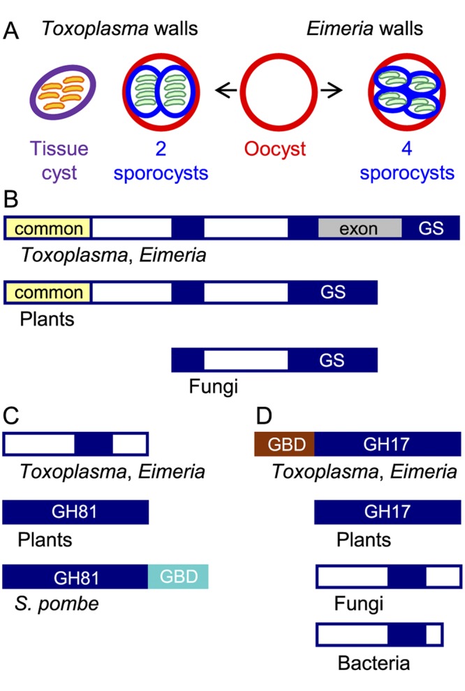
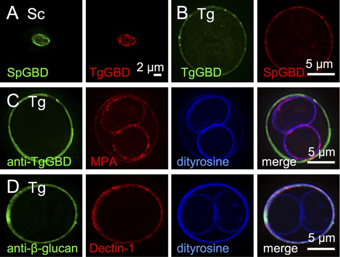
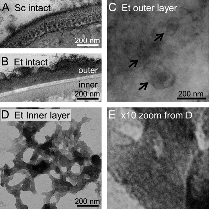
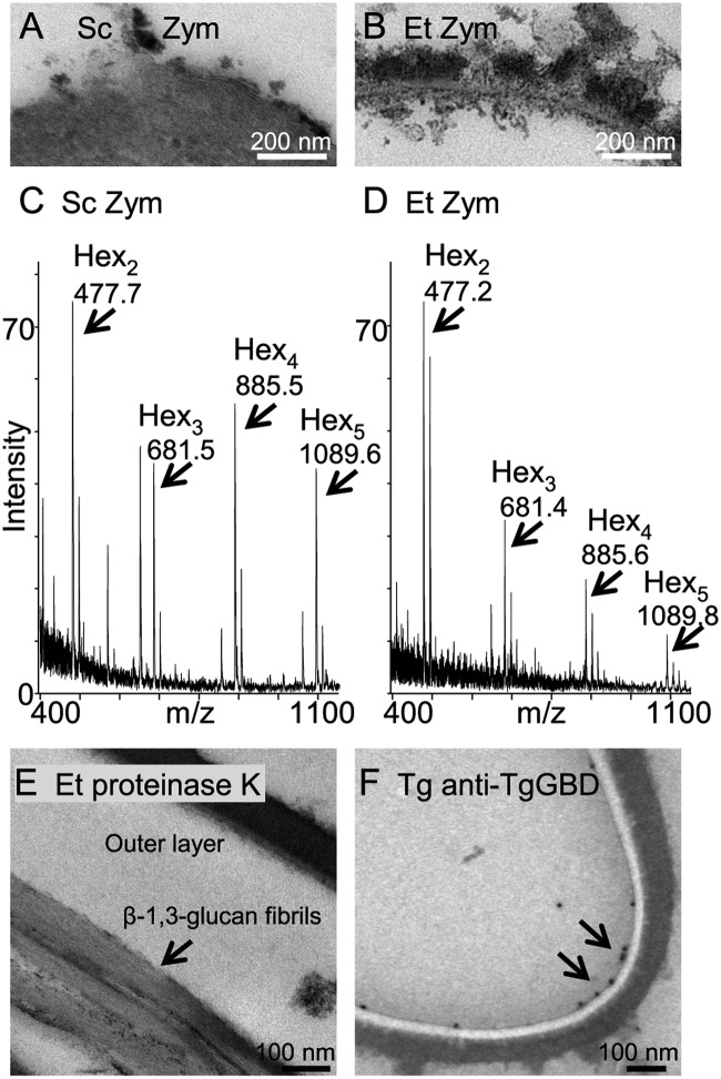
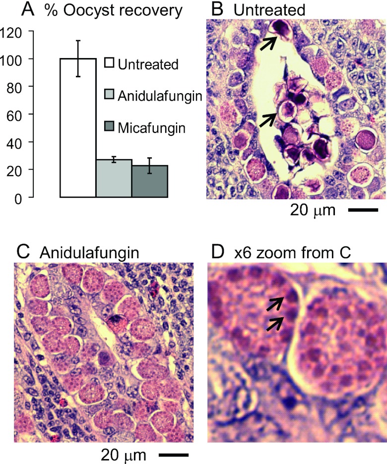
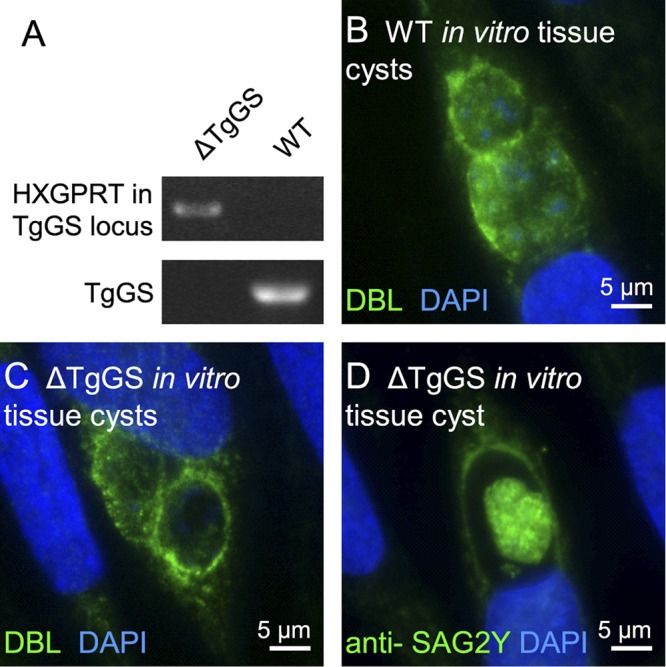
The walls of infectious pathogens, which are essential for transmission, pathogenesis, and diagnosis, contain sugar polymers that are defining structural features, e.g., β-1,3-glucan and chitin in fungi, chitin in Entamoeba cysts, β-1,3-GalNAc in Giardia cysts, and peptidoglycans in bacteria. The goal here was to determine in which of three walled forms of Toxoplasma gondii (oocyst, sporocyst, or tissue cyst) is β-1,3-glucan, the product of glucan synthases and glucan hydrolases predicted by whole-genome sequences of the parasite. The three most important discoveries were as follows. (i) β-1,3-glucan is present in oocyst walls of Toxoplasma and Eimeria (a chicken parasite that is a model for intestinal stages of Toxoplasma) but is absent from sporocyst and tissue cyst walls. (ii) Fibrils of β-1,3-glucan are part of a trabecular scaffold in the inner layer of the oocyst wall, which also includes a glucan hydrolase that has a novel glucan-binding domain. (iii) Echinocandins, which target the glucan synthase and kill fungi, arrest development of the Eimeria oocyst wall and prevent release of the parasites into the intestinal lumen. In summary, β-1,3-glucan, which can be targeted by drugs, is an important component of oocyst walls of Toxoplasma but is not a component of sporocyst and tissue cyst walls.

Importance: We show here that walls of Toxoplasma oocysts, the infectious stage shed by cats, contain β-1,3-glucan, a sugar polymer that is a major component of fungal walls. In contrast to fungi, β-1,3-glucan is part of a trabecular scaffold in the inner layer of the oocyst wall that is independent of the permeability barrier formed by the outer layer of the wall. While glucan synthase inhibitors kill fungi, these inhibitors arrest the development of the oocyst walls of Eimeria (an important chicken pathogen that is a surrogate for Toxoplasma) and block release of oocysts into the intestinal lumen. The absence of β-1,3-glucan in tissue cysts of Toxoplasma suggests that drugs targeted at the glucan synthase might be used to treat Eimeria in chickens but not to treat Toxoplasma in people.


Figures

FIG 1
FIG 1
The glucan synthase and one glucan hydrolase of Toxoplasma gondii and Eimeria tenella resemble plant rather than fungal enzymes. (A) Toxoplasma has three walled forms: cat shed oocysts (red wall) in their feces, which sporulate in the environment to form two internal sporocysts (blue wall), each with four sporozoites (green). Tissue cysts (purple wall) in the muscle or brain of any warm-blooded animal contain numerous bradyzoites (orange). Chickens shed Eimeria oocysts in their feces that sporulate to form four sporocysts, each of which contains two sporozoites. (B) Toxoplasma and Eimeria glucan synthases have a conserved glucan synthase (GS) domain that is split into three parts (GS in blue), a domain common to plants, but not to fungi (yellow), and an exon (gray) present only in parasite glucan synthases. The white domains are unique to parasites, plants, or fungi. (C) One Toxoplasma and Eimeria glucan hydrolase contains a portion of the GH81 glycohydrolase domain present in plant and fungal enzymes but does not have a C-terminal glucan-binding domain (GBD in turquoise) present in the Schizosaccharomyces pombe (Eng1) glucan hydrolase. (D) The other Toxoplasma and Eimeria glucan hydrolase contains a GH17 glycohydrolase domain similar to that of plants and different from those of fungi and bacteria. The parasite glucan hydrolase also contains a glucan-binding domain (GBD in brown) between the signal peptide and the GH17 domain. The lectin activity of the TgGBD, which is not phylogenetically related to the SpGBD, is shown in Fig. 2. See Fig. S1 in the supplemental material for sequences of GBD and GH17 domains of the Toxoplasma glucan hydrolase.
FIG 2
FIG 2
Oocyst walls, but not sporocyst walls, of Toxoplasma gondii are labeled with β-1,3-glucan-binding reagents. (A) Walls of Saccharomyces cerevisiae (Sc), deproteinated with NaOH, bind glucan-binding domains from Schizosaccaromyces pombe (SpGBD in green) and Toxoplasma (TgGBD in red) glucan hydrolases, which have been expressed as MBP (maltose-binding protein) fusion proteins and labeled with Alexa Fluor dyes. (B) SpGBD and TgGBD also bind to Toxoplasma gondii (Tg) oocyst walls, but not to sporocyst walls. MBP controls fail to bind to yeast or parasite walls. (C) Antibodies to the Toxoplasma glucan-binding domain (anti-TgGBD in green) bind to oocyst, but not sporocysts, walls of frozen and thawed Toxoplasma. For a control for permeability of sporulated oocysts, the plant lectin MPA (red) that binds to GalNAc labels both the oocyst and sporocyst walls, which are autofluorescent in the UV channel due to the presence of dityrosines. (D) Toxoplasma oocyst walls also are labeled by antibodies to β-1,3-glucan (green) and by the macrophage lectin Dectin-1 (red). See Fig. S2 in the supplemental materialfor fluorescent micrographs of Eimeria oocysts labeled with the same glucan-binding reagents.
FIG 3
FIG 3
The inner layer of the oocyst wall of Eimeria is a trabecular scaffold. (A) Transmission electron microscopy of cross sections stained with ruthenium red shows that the intact Saccharomyces cerevisiae (Sc) wall is one continuous structure. (B) The Eimeria tenella (Et) oocyst wall is made of an electron-dense outer layer, an electron-lucent interlayer, and a moderately electron-dense inner layer. (C) Negative staining of intact oocysts with ruthenium red shows that the outer layer of the Eimeria oocyst wall is relatively smooth with occasional oval objects that vary in size (arrows). (D) The inner layer of the Eimeria oocyst wall, which is revealed by negatively staining walls that have been mechanically disrupted, is a trabecular scaffold. (E) High-power view of panel D shows putative fibrils of β-1,3-glucan, which are straight and present in parallel arrays, within the trabecular scaffolds. The inner layer of oocyst walls of Toxoplasma are also composed of similar trabecular scaffolds.
FIG 4
FIG 4
Zymolyase releases glucose oligomers from the oocyst walls of Eimeria. (A) Zymolyase (Zym), which is a mixture of β-glucanases and proteases from Arthrobacter luteus, completely removes the walls of Saccharomyces cerevisiae (Sc) to form spheroplasts. (B) In contrast, zymolyase primarily removes the inner layer of oocyst walls of Eimeria. (C) MALDI-TOF MS of glucose oligomers released by zymolyase from Saccharomyces walls. The smaller peaks to the right of each peak marked with arrows are potassium adducts. A peak at 647.6 is unidentified. (D) MALDI-TOF MS of glucose oligomers released by zymolyase from Eimeria oocyst walls are similar to those of Saccharomyces. There are no oligosaccharides present in control samples in which zymolyase was omitted. (E) Proteinase K has little effect on the outer layer of oocyst walls of Eimeria but occasionally releases bundles of fibrils (presumably composed of β-1,3-glucan) (arrow) from the inner layer of the oocyst walls. (F) Antibodies to the Toxoplasma glucan-binding domain (anti-TgGBD antibodies visualized with immunogold) (arrows) bind to the inner layer of the Toxoplasma oocyst wall that is weakly stained in the absence of ruthenium red.
FIG 5
FIG 5
Echinocandins arrest the development of oocyst walls of Eimeria in the ceca of infected chickens. (A) Oocyst recoveries are markedly reduced versus untreated chickens in chickens treated with 10 mg/kg anidulafungin or 10 mg/kg micafungin. Error bars show standard deviations of the oocyst counts in two experiments in which there were two chickens per group. (B) Hematoxylin-and-eosin-stained section of the cecum of an untreated chicken infected with Eimeria shows zygotes in numerous developmental stages, including some with small secretory vesicles (early), large secretory vesicles near the periphery (later), and darkly stained walls (fully developed and often released into the lumen of the crypt) (arrows). (C) A cecum from a chicken treated with anidulafungin shows numerous zygotes that are arrested, so few walled forms are present. Similar results were observed with micafungin. (D) High-power view of panel C shows large purple secretory vesicles at the periphery of developing oocysts arrested by anidulafungin (arrows).
FIG 6
FIG 6
Knockout experiments show that glucan synthase is not essential for tissue cyst formation by Toxoplasma in vitro. (A) PCR products for the HXGPRT gene in the glucan synthase locus are present in the glucan synthase knockout (ΔTgGS), but not in nontransformed Toxoplasma (wild type [WT]). Conversely, the Toxoplasma glucan synthase gene (TgGS) is present in nontransformed parasites, but not in the ΔTgGS parasites. (B) In vitro tissue cysts from nontransformed Toxoplasma are labeled with DBL (green walls), while nuclei are labeled with DAPI (blue). (C) ΔTgGS in vitro tissue cysts showlabeling of walls with DBL similar to that of nontransformed tissue cysts shown in panel B, (D) ΔTgGS bradyzoites within an in vitro tissue cyst are la beled with anti-SAG2Y antibody similarly to nontransformed bradyzoites.

References

    1. Boothroyd JC. 2009. Toxoplasma gondii: 25 years and 25 major advances for the field. Int. J. Parasitol. 39:935–946 - PMC - PubMed
    1. Weiss LM, Dubey JP. 2009. Toxoplasmosis: a history of clinical observations. Int. J. Parasitol. 39:895–901 - PMC - PubMed
    1. Dubey JP, Lindsay DS, Speer CA. 1998. Structures of Toxoplasma gondii tachyzoites, bradyzoites, and sporozoites and biology and development of tissue cysts. Clin. Microbiol. Rev. 11:267–299 - PMC - PubMed
    1. Belli SI, Smith NC, Ferguson DJ. 2006. The coccidian oocyst: a tough nut to crack! Trends Parasitol. 22:416–423 - PubMed
    1. Ferguson DJ, Belli SI, Smith NC, Wallach MG. 2003. The development of the macrogamete and oocyst wall in Eimeria maxima: immuno-light and electron microscopy. Int. J. Parasitol. 33:1329–1340 - PubMed

Publication types

LinkOut - more resources