. 2012 Sep 25;3(5):e00279-12.
doi: 10.1128/mBio.00279-12. Print 2012.

Propionibacterium acnes bacteriophages display limited genetic diversity and broad killing activity against bacterial skin isolates

Affiliations

Propionibacterium acnes bacteriophages display limited genetic diversity and broad killing activity against bacterial skin isolates

Laura J Marinelli et al. mBio. .

Abstract

Investigation of the human microbiome has revealed diverse and complex microbial communities at distinct anatomic sites. The microbiome of the human sebaceous follicle provides a tractable model in which to study its dominant bacterial inhabitant, Propionibacterium acnes, which is thought to contribute to the pathogenesis of the human disease acne. To explore the diversity of the bacteriophages that infect P. acnes, 11 P. acnes phages were isolated from the sebaceous follicles of donors with healthy skin or acne and their genomes were sequenced. Comparative genomic analysis of the P. acnes phage population, which spans a 30-year temporal period and a broad geographic range, reveals striking similarity in terms of genome length, percent GC content, nucleotide identity (>85%), and gene content. This was unexpected, given the far-ranging diversity observed in virtually all other phage populations. Although the P. acnes phages display a broad host range against clinical isolates of P. acnes, two bacterial isolates were resistant to many of these phages. Moreover, the patterns of phage resistance correlate closely with the presence of clustered regularly interspaced short palindromic repeat elements in the bacteria that target a specific subset of phages, conferring a system of prokaryotic innate immunity. The limited diversity of the P. acnes bacteriophages, which may relate to the unique evolutionary constraints imposed by the lipid-rich anaerobic environment in which their bacterial hosts reside, points to the potential utility of phage-based antimicrobial therapy for acne.

Importance: Propionibacterium acnes is a dominant member of the skin microflora and has also been implicated in the pathogenesis of acne; however, little is known about the bacteriophages that coexist with and infect this bacterium. Here we present the novel genome sequences of 11 P. acnes phages, thereby substantially increasing the amount of available genomic information about this phage population. Surprisingly, we find that, unlike other well-studied bacteriophages, P. acnes phages are highly homogeneous and show a striking lack of genetic diversity, which is perhaps related to their unique and restricted habitat. They also share a broad ability to kill clinical isolates of P. acnes; phage resistance is not prevalent, but when detected, it appears to be conferred by chromosomally encoded immunity elements within the host genome. We believe that these phages display numerous features that would make them ideal candidates for the development of a phage-based therapy for acne.


Figures

FIG 1
FIG 1
P. acnes phage virion morphologies. Negatively stained electron micrographs of the 11 P. acnes phages characterized in this study reveal that all have the same siphoviral virion morphotype. All of these phages have similarly sized isometric heads (~50 nm in diameter) and long, flexible tails (~150 nm in length).
FIG 2
FIG 2
Percent GC contents of bacteriophages and their hosts. The percent GC contents of the genomes of individual bacteriophages that infect P. acnes, M. smegmatis, Staphylococcus sp., or Pseudomonas sp. are plotted in order, from the lowest value to the highest. The analysis includes 14 phages of P. acnes, 221 phages of M. smegmatis, 66 phages of Staphylococcus sp., and 51 phages of Pseudomonas sp. (see Materials and Methods). The percent GC contents of host bacteria and their close relatives, which may serve as potential hosts, were calculated from published genomes; these are shown as dotted lines and listed with percent GC contents in parentheses.
FIG 3
FIG 3
Dot plot nucleotide sequence comparisons of P. acnes phages, mycobacteriophages, Staphylococcus phages, and Pseudomonas phages. The genome sequences of 14 P. acnes phages, 221 mycobacteriophages, 66 Staphylococcus phages, and 51 Pseudomonas phages were concatenated to form four compiled sequences of 0.41, 15.46, 3.3, and 3.88 Mbp, respectively. Each was then compared with itself using the dot plot program Gepard (48).
FIG 4
FIG 4
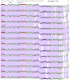
Whole-genome comparisons of P. acnes phages. The genome maps of the 14 completely sequenced P. acnes phages are shown with the pairwise nucleotide sequence similarities displayed as colored segments between the genomes; the strength of sequence similarity is represented according to a color spectrum in which violet is the most similar and red is the least. The positions of predicted genes are shown as boxes either above (transcribed rightward) or below (transcribed leftward) each genome, with gene numbers shown within the boxes; putative gene functions are noted at the top. The maps were generated using the program Phamerator (49) and a database named acnes_myco30 (see text).
FIG 5
FIG 5
Organization of the P100D genome and its percent GC content variation. A genome map of P. acnes phage P100D is shown with predicted genes represented as colored boxes either above (transcribed rightward) or below (transcribed leftward) the genome. The gene number is shown within each box, and the “phamily” to which that the gene corresponds is displayed above, with the number of “phamily” members shown in parentheses. Putative gene functions are noted with arrows indicating the orientation of transcription. All of the other genomes described here have the same basic organization. At the bottom is a scan of percent GC contents across the P100D genome done with window and step sizes of 100 and 50 bp, respectively. The sharp deviation of percent GC content at the right end of the genome occurs in all of the P. acnes phages.
FIG 6
FIG 6
Gene content relationships between P. acnes phages and phages of related hosts. (A) The relationships between the 14 P. acnes phages and a selection of phages that infect other bacteria of the order Actinomycetales were obtained by using the NeighborNet function in Splitstree4 (51). The P. acnes, Mycobacterium, Rhodococcus, Corynebacterium. Streptomyces, Tsukamurella, and Gordonia phages are shown in the red, green, orange, blue, yellow, aqua, and mauve circles, respectively. (B) Detailed view of the display in panel A showing the Pacnes phages.
FIG 7
FIG 7
Organization of P. acnes phage endolysins. (A) Organization of the P. acnes endolysins and their closest homologues present in Rhodococcus phages ReqiDocB7 and REQ2 and a group of subcluster B1 mycobacteriophages of which KLucky39 is representative. All have an amidase-2 domain that cleaves peptidoglycan but have different and unrelated C-terminal domains, which are presumed to confer cell wall binding specificity. The KLucky39 endolysin—like many of the mycobacteriophage endolysins—contains three domains, including an N-terminal M23 peptidase (26). (B) Alignment of the P. acnes phage endolysins shows that they are closely related and display the greatest amino acid sequence differences at around position 200, which likely corresponds to a glycine-rich interdomain linker.
FIG 8
FIG 8
Host preferences of P. acnes phages. The efficiency of plating of each of the 11 P. acnes phages was calculated for 27 bacterial isolates, including 23 donor isolates and four ATCC strains; ATCC 6919 was used as a reference for normalization. Eleven isolates and ATCC 11827 have variable degrees of phage sensitivity; most differences result from modest reductions in plating efficiency (10- to 1,000-fold relative to reference strain ATCC 6919), but two isolates, B66.8 and B101.9, are strongly resistant to 9 and 10 of the phages, respectively (plating efficiencies are reduced by at least 1,000,000-fold). The remaining 12 isolates and three ATCC strains are infected equivalently by all of the phages.
FIG 9
FIG 9
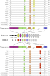
Correspondence of CRISPR spacer sequences and P. acnes phages. Pacnes strains B101.9 and B66.8 each contain a CRISPR locus containing at least the cas3 and csc4 genes flanked by an array of direct repeats separated by 33-bp variable spacer sequences. The spacers are represented by colored boxes, and those numbered 3, 4, and 5 are identical in the two strains. Each of the first five spacers has closely related sequences in the P. acnes phages, with the corresponding genes indicated. The numbers of nucleotide sequence deviations from each of the 33-bp spacers in the 14 P. acnes phage genomes are represented above (for strain 101.9) and below (strain B66.8) the CRISPRs. Those phage segments that have two or fewer sequence departures from the spacer are boxed in yellow. At the right, the patterns of phage resistance (R) or sensitivity (S) to the two strains are indicated; NT, not tested.

References

    1. Casjens SR. 2008. Diversity among the tailed-bacteriophages that infect the Enterobacteriaceae. Res. Microbiol. 159:340–348 - PMC - PubMed
    1. Hatfull GF, Science Education Alliance Phage Hunters Advancing Genomics and Evolutionary Science Program, KwaZulu-Natal Research Institute for Tuberculosis and HIV Mycobacterial Genetics Course Students, Phage Hunters Integrating Research and Education Program. 2012. Complete genome sequences of 138 mycobacteriophages. J. Virol. 86:2382–2384 - PMC - PubMed
    1. Hatfull GF, et al. 2010. Comparative genomic analysis of sixty mycobacteriophage genomes: genome clustering, gene acquisition, and gene size. J. Mol. Biol. 397:119–143 - PMC - PubMed
    1. Pope WH, et al. 2011. Cluster K mycobacteriophages: insights into the evolutionary origins of mycobacteriophage TM4. PLoS One 6:e26750 - PMC - PubMed
    1. Pope WH, et al. 2011. Expanding the diversity of mycobacteriophages: insights into genome architecture and evolution. PLoS One 6:e16329. - PMC - PubMed

Publication types

MeSH terms