. 2012 Nov;23(22):4465-71.
doi: 10.1091/mbc.E12-09-0655. Epub 2012 Sep 26.

Displacement of basolateral Bazooka/PAR-3 by regulated transport and dispersion during epithelial polarization in Drosophila

Affiliations

Displacement of basolateral Bazooka/PAR-3 by regulated transport and dispersion during epithelial polarization in Drosophila

R F Andrew McKinley et al. Mol Biol Cell. 2012 Nov.

Abstract

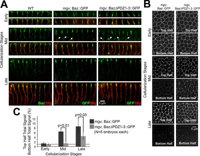
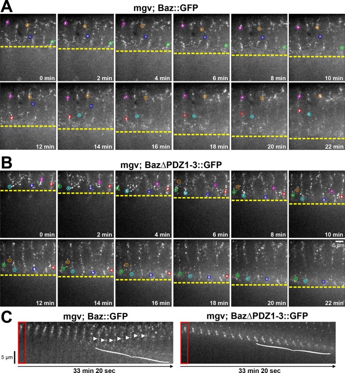
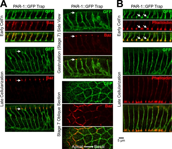
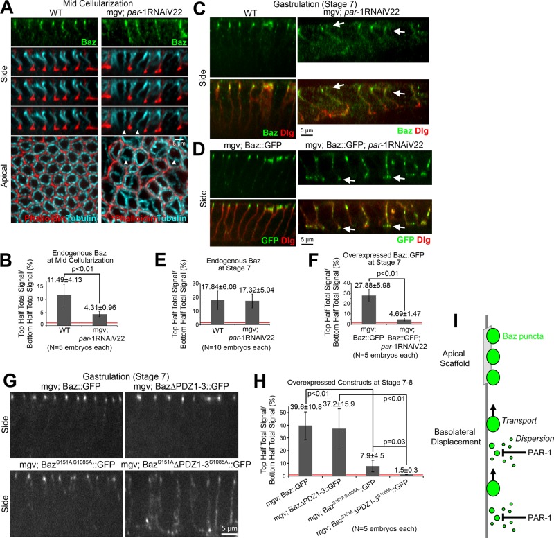
Polarity landmarks guide epithelial development. In the early Drosophila ectoderm, the scaffold protein Bazooka (Drosophila PAR-3) forms apicolateral landmarks to direct adherens junction assembly. However, it is unclear how Bazooka becomes polarized. We report two mechanisms acting in concert to displace Bazooka from the basolateral membrane. As cells form during cellularization, basally localized Bazooka undergoes basal-to-apical transport. Bazooka requires its three postsynaptic density 95, discs large, zonula occludens-1 (PDZ) domains to engage the transport mechanism, but with the PDZ domains deleted, basolateral displacement still occurs by gastrulation. Basolateral PAR-1 activity appears to act redundantly with the transport mechanism. Knockdown of PAR-1 sporadically destabilizes cellularization furrows, but basolateral displacement of Bazooka still occurs by gastrulation. In contrast, basolateral Bazooka displacement is blocked with disruption of both the transport mechanism and phosphorylation by PAR-1. Thus Bazooka is polarized through a combination of transport and PAR-1-induced dispersion from basolateral membranes. Our work complements recent findings in Caenorhabditis elegans and thus suggests the coupling of transport and dispersion is a common protein polarization strategy.


Figures

FIGURE 1:
FIGURE 1:
The PDZ domains of Baz promote its apical enrichment during cellularization. (A) Fixed early (5- to 7-μm furrows), mid (12- to 14-μm furrows), and late (19- to 21-μm furrows) cellularization embryos. Endogenous Baz vs. Baz::GFP and Baz∆PDZ1-3::GFP expressed maternally with mgv. Discs large (Dlg) shows furrows. Arrows show basal Baz puncta. (B) Projections of top and bottom halves of lateral membranes. (C) Total fluorescence intensity ratios of top vs. bottom halves of lateral membranes. Red line shows 1:1 top:bottom ratio.
FIGURE 2:
FIGURE 2:
The PDZ domains of Baz are required for its basal-to-apical transport. (A, B) Cellularizing embryos over 22 min. Single confocal planes of central cross sections. Circles track puncta in plane for extended periods. Constructs expressed maternally with mgv. (A) As furrow canals progress (yellow line), Baz::GFP puncta stay apical (pink, orange, and blue circles), move basally (green circles), or translocate basally to apically (red and cyan circles). (B) As furrow canals progress (yellow line), Baz∆PDZ1-3::GFP puncta move basally with them (cyan, blue, red, green, pink, and orange circles) without apical translocations. (C) Kymographs of cellularizing embryo cross sections. Baz::GFP puncta translocate apically (arrowheads). Basal Baz::GFP and Baz∆PDZ1-3::GFP puncta disperse at late cellularization (brackets; apical puncta lack photobleaching).
FIGURE 3:
FIGURE 3:
PAR-1 segregates from apicolateral Baz by gastrulation. (A) Fixed PAR-1::GFP trap embryos. PAR-1 segregates below endogenous Baz beginning at late cellularization (arrows). Oblique stage 7 section shows Baz above PAR-1. (B) PAR-1::GFP trap embryos stained with phalloidin. Note PAR-1 and actin furrow canal coenrichment at early cellularization (arrows).
FIGURE 4:
FIGURE 4:
PAR-1 phosphorylation contributes redundantly to Baz polarization. (A) Fixed midcellularization embryos. In WT, Baz localizes apicolaterally, phalloidin stains overall furrows and actin-rich furrow canals, and apical-basal microtubule bundles form baskets around nuclei. With maternal expression of par-1 shRNA (Valium22 line) with mgv, Baz mislocalizes basally and furrows are lost sporadically (arrowheads), but microtubule baskets are present. (B) Total fluorescence intensity ratios of endogenous Baz puncta in the top vs. bottom halves of lateral membranes. (C, D) Fixed stage 7 embryos. (C) With and without PAR-1 RNAi, endogenous Baz is apicolaterally enriched despite PAR-1 RNAi morphological defects. Dlg shows basolateral membranes. (D) Insertion of UAS-Baz::GFP at attp40 (chromosome 2) allowed maternal coexpression with par-1 shRNA (Valium22 line) at attp2 (chromosome 3; other Baz constructs at attp2). With coexpression, Baz::GFP mislocalized basally, accumulating at the base of lateral membranes (arrows). (E, F) Total fluorescence intensity ratios of puncta in the top vs. bottom halves of cells. (G) Fixed stage 7 embryo side views with Baz::GFP, Baz∆PDZ1-3::GFP, BazS151AS1085A::GFP, and BazS151A∆PDZ1-3S1085A::GFP expressed maternally with mgv. (H) Total fluorescence intensity ratios of protein puncta in top vs. bottom halves of cells. Red lines show 1:1 top:bottom ratios. (I) Model of Baz polarization.

Similar articles

Cited by

References

    1. Bayraktar J, Zygmunt D, Carthew RW. Par-1 kinase establishes cell polarity and functions in Notch signaling in the Drosophila embryo. J Cell Sci. 2006;119:711–721. - PubMed
    1. Benton R, St Johnston D. A conserved oligomerization domain in Drosophila Bazooka/PAR-3 is important for apical localization and epithelial polarity. Curr Biol. 2003a;13:1330–1334. - PubMed
    1. Benton R, St Johnston D. Drosophila PAR-1 and 14-3-3 inhibit Bazooka/PAR-3 to establish complementary cortical domains in polarized cells. Cell. 2003b;115:691–704. - PubMed
    1. Bohm H, Brinkmann V, Drab M, Henske A, Kurzchalia TV. Mammalian homologues of C. elegans PAR-1 are asymmetrically localized in epithelial cells and may influence their polarity. Curr Biol. 1997;7:603–606. - PubMed
    1. Doerflinger H, Benton R, Shulman JM, St Johnston D. The role of PAR-1 in regulating the polarised microtubule cytoskeleton in the Drosophila follicular epithelium. Development. 2003;130:3965–3975. - PubMed

Publication types

MeSH terms

Substances

LinkOut - more resources