

doi: 10.1177/1941738109350438.
The basic science of articular cartilage: structure, composition, and function
Affiliations
- PMID: 23015907
- PMCID: PMC3445147
- DOI: 10.1177/1941738109350438
Item in Clipboard
The basic science of articular cartilage: structure, composition, and function
Sports Health.
2009 Nov.
No abstract available
Keywords: articular cartilage; basic science; chondrocyte; collagen; extracellular matrix; proteoglycan.
Conflict of interest statement
No potential conflict of interest declared.
Figures
Gross photograph of healthy articular cartilage in an adult human knee.
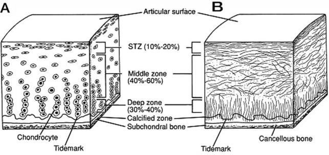
Schematic, cross-sectional diagram of healthy articular cartilage: A, cellular organization in the zones of articular cartilage; B, collagen fiber architecture. (Copyright American Academy of Orthopaedic Surgeons. Reprinted from the Journal of the American Academy of Orthopaedic Surgeons, 1994;2:192-201 with permission.)
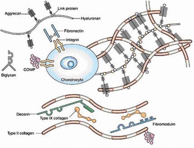
Extracellular matrix of articular cartilage. Two major load-bearing macromolecules are present in articular cartilage: collagens (mainly, type II) and proteoglycans (notably, aggrecan). Smaller classes of molecules, such as noncollagenous proteins and smaller proteoglycans, are present in smaller amounts. The interaction between the highly negatively charged cartilage proteoglycans and type II collagen provides the compressive and tensile strength of the tissue. (Reprinted with permission from Chen et al, 2006.)
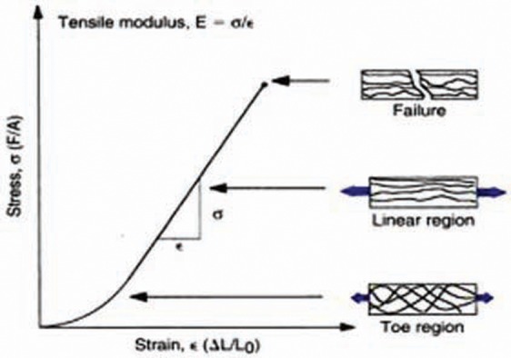
A stress-strain diagram for articular cartilage during tensile loading. The schematic representations on the right illustrate the orientation of the collagen fibrils in response to loading. Reprinted with permission from Nordin and Frankel, Basic Biomechanics of the Musculoskeletal System.
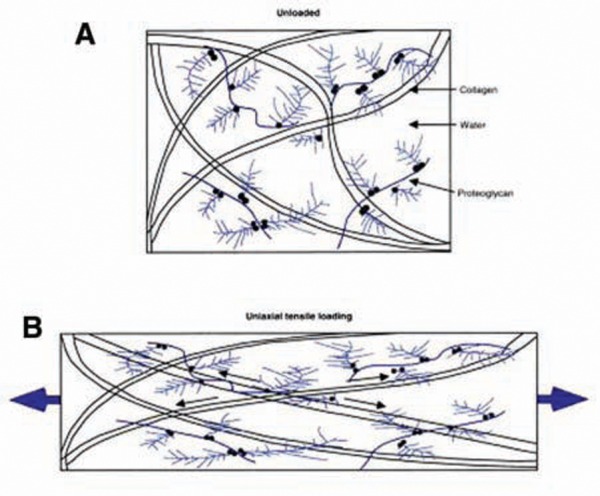
A schematic depiction of the main components of articular cartilage when the tissue is unloaded (A) and when tensile load is applied (B). When the tissue is loaded, collagen fibrils align along the axis of tension. Reprinted with permission from Nordin and Frankel, Basic Biomechanics of the Musculoskeletal System.,
References
-
- Akizuki S, Mow VC, Müller F, Pita JC, Howell DS, Manicourt DH. Tensile properties of human knee joint cartilage: I. Influence of ionic cartilage conditions, weight bearing and fibrillation on the tensile modulus. J Orthop Res. 1986;4(4):379-392 - PubMed
-
- Alford JW, Cole BJ. Cartilage restoration, part I: basic science, historical perspective, patient evaluation and treatment options. Am J Sports Med. 2005;33:295-306 - PubMed
-
- Ateshian GA, Warden WH, Kim JJ, et al. Finite deformation biphasic material properties of bovine articular cartilage from confined compression experiments. J Biomech. 1997;30:1157-1164 - PubMed
-
- Bashir A, Gray ML, Boutin RD, Burstein D. Glycosaminoglycan in articular cartilage: in vivo assessment with delayed Gd(DPTA)(2-)-enhanced MR imaging. Radiology. 1997;205:551-558 - PubMed
-
- Bashir A, Gray ML, Burstein D. Gd-DPTA2- as a measure of cartilage degradation. Magn Reson Med. 1996;36:665-673 - PubMed
LinkOut - more resources
Full Text Sources
Other Literature Sources
Medical
