. 2010 Sep;2(5):403-9.
doi: 10.1177/1941738110378987.

Femoroacetabular Impingement in Athletes, Part II: Treatment and Outcomes

Affiliations

Femoroacetabular Impingement in Athletes, Part II: Treatment and Outcomes

J W Thomas Byrd. Sports Health. 2010 Sep.

Abstract

Context: Femoroacetabular impingement (FAI) is a common cause of hip pathology and secondary dysfunction among athletes. Much information has been gained regarding the cause and pathomechanics of this disorder. Now, efforts are focusing on treatment to restore the joint and reduce the secondary damage that causes painful dysfunction.

Evidence acquisition: This article reviews the scientific literature in reference to treatment of FAI in athletes.

Results: Several studies reported reasonably successful outcomes in the arthroscopic management of FAI in athletes, and 1 study reported on open surgical correction of this disorder. Few major complications have been described.

Conclusions: When the diagnosis is given early, some athletes may benefit from a rehabilitation strategy that includes training modifications to protect the at-risk hip. When indicated, arthroscopic surgery can address the joint damage and correct the underlying impingement. Although the joint may not be normal, successful results with return to sports can often be expected.

Keywords: assessment; athletes; etiology; femoroacetabular impingement; hip arthroscopy.


Conflict of interest statement

One or more authors has declared a potential conflict of interest: J. W. Thomas Byrd is a consultant for Smith & Nephew and A2 Surgical and has received research funding from Smith & Nephew.

Figures

Figure 1.
Figure 1.
A, the anterior portal coincides with the intersection of a sagittal line drawn distally from the anterior superior iliac spine and a transverse line across the superior margin of the greater trochanter. The portal courses approximately 45° cephalad and 30° toward the midline. The anterolateral and posterolateral portals are positioned directly over the superior aspect of the trochanter at the anterior and posterior borders. B, the femoral artery and nerve lie well medial to the anterior portal. The sciatic nerve lies posterior to the posterolateral portal. The lateral femoral cutaneous nerve lies close to the anterior portal.
Figure 2.
Figure 2.
A 38-year-old woman with progressive pain and loss of motion of the right hip. A, 3-dimensional computed tomography scan illustrates pincer impingement (arrows) and a kissing lesion characterized by osteophyte formation on the femoral head (asterisk). B, as viewed from the anterolateral portal, there is maceration of the anterior labrum (white asterisk) and associated articular delamination (black asterisk). C, debridement of the degenerate labrum exposes the pincer lesion (arrows). D, the pincer lesion is recontoured with a burr. E, a postoperative 3-dimensional computed tomography scan demonstrates the extent of bony recontouring of the acetabulum and the femoral head.
Figure 3.
Figure 3.
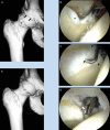
A 15-year-old female gymnast with pain and reduced internal rotation of the left hip. A, a 3-dimensional computed tomography scan defines a pincer lesion with accompanying os acetabulum (arrow) and cam lesion (asterisk). B, as viewed from the anterolateral portal, the pincer lesion and os acetabulum (asterisk) are exposed, with the labrum being sharply released with an arthroscopic knife. C, the acetabular fragment has been removed and the rim trimmed with anchors placed to repair the labrum. D, the labrum has been refixed.
Figure 4.
Figure 4.
A 20-year-old hockey player with a 4-year history of right hip pain. A, a 3-dimensional computed tomography scan defines the cam lesion (arrows). B, as viewed from the anterolateral portal, the probe introduced anteriorly displaces an area of articular delamination from the anterolateral acetabulum characteristic of the peel-back phenomenon created by the bony lesion shearing the articular surface during hip flexion.
Figure 5.
Figure 5.
A, a capsulotomy performed by connecting the anterior and anterolateral portals is necessary for the instruments to pass freely from the central to peripheral compartment as the traction is released and the hip flexed. B, with the hip flexed, the anterolateral portal is now positioned along the neck of the femur. A cephalad (proximal) anterolateral portal has been placed, and the original anterior and posterolateral portals have been removed. These 2 portals allow access to the entirety of the cam lesion in most cases. Their position also allows an unhindered view with the c-arm.
Figure 6.
Figure 6.
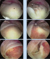
View from the periphery. A, a cam lesion covered with fibrocartilage (asterisk). B, an arthroscopic curette used to denude the abnormal bone. C, excision area is fully exposed. D, bony resection at the articular margin. E, the completed recontouring. F, lateral view on the base of the neck; the lateral retinacular vessels identified (arrows) and preserved.

References

    1. Bizzini M, Notzli HIP, Maffiuletti NA. Femoroacetabular impingement in professional ice hockey players. a case series of five athletes after open surgical decompression of the hip. Am J Sports Med. 2007;35:1955. - PubMed
    1. Byrd JWT. Hip arthroscopy by the supine approach. Instr Course Lect. 2006;55:325-336 - PubMed
    1. Byrd JWT. Hip arthroscopy utilizing the supine position. Arthroscopy. 1994;10:275-280 - PubMed
    1. Byrd JWT. The supine approach. In: Byrd JWT, ed. Operative Hip Arthroscopy. 2nd ed. New York, NY: Springer; 2005:145-169
    1. Byrd JWT, Jones KS. Arthroscopic management of femoroacetabular impingement. Instr Course Lect. 2009;58:231-239 - PubMed