. 2012 May;4(3):236-45.
doi: 10.1177/1941738112442484.

Rowing injuries

Affiliations

Rowing injuries

Timothy M Hosea et al. Sports Health. 2012 May.

Abstract

Context: Rowing is one of the original modern Olympic sports and was one of the most popular spectator sports in the United States. Its popularity has been increasing since the enactment of Title IX. The injury patterns in this sport are unique because of the stress applied during the rowing stroke.

Evidence acquisition: This review summarizes the existing literature describing the biomechanics of the rowing stroke and rowing-related injury patterns. Data were obtained from previously published peer-reviewed literature through a search of the entire PubMed database (up to December, 2011) as well as from textbook chapters and rowing coaching manuals.

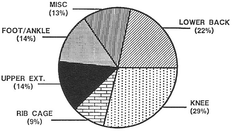
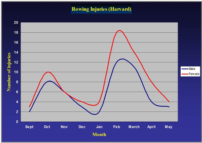
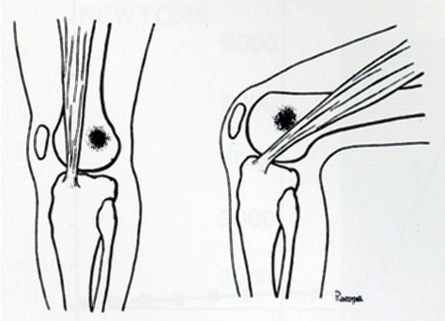
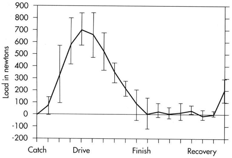
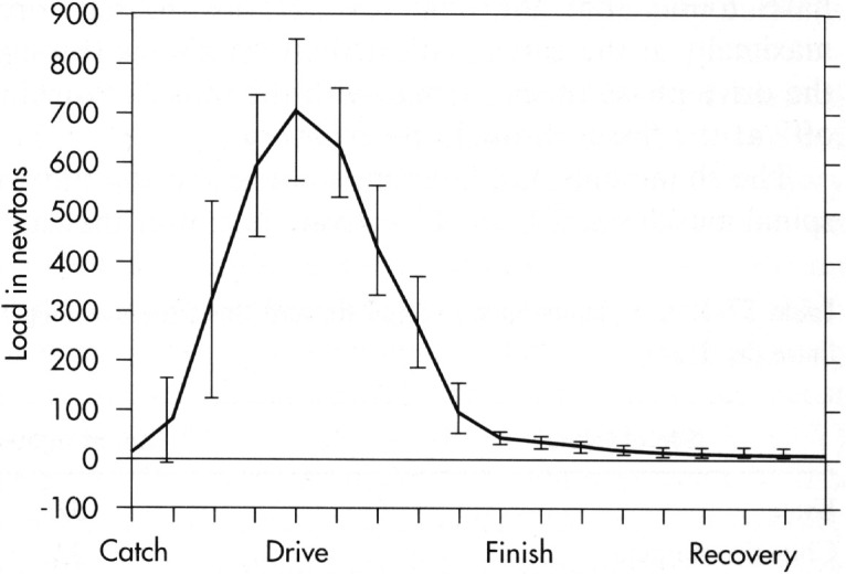
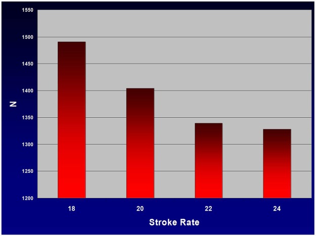
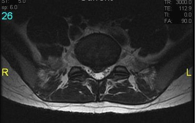
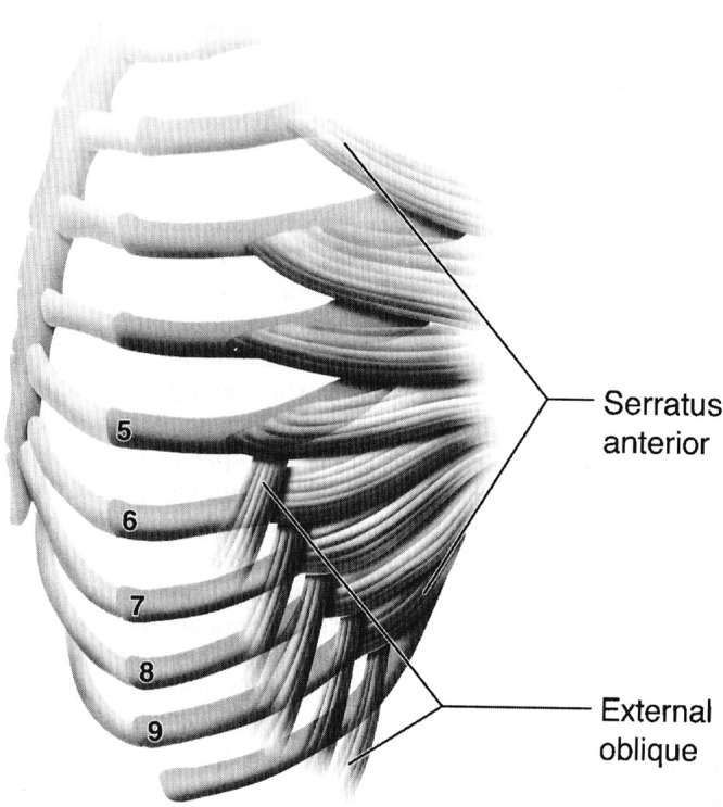
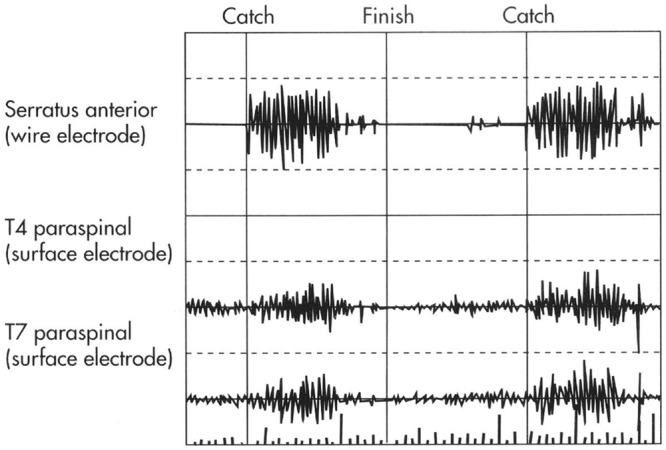
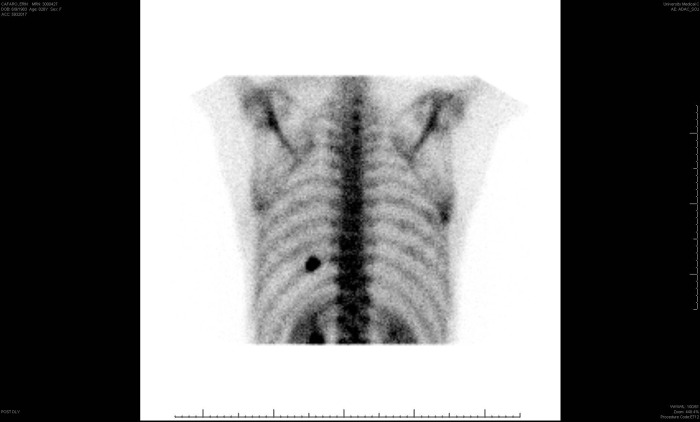
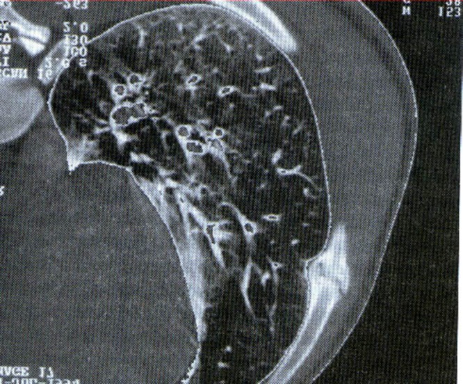
Results: Rowing injuries are primarily overuse related. The knee, lumbar spine, and ribs are most commonly affected. The injury incidence is directly related to the volume of training and technique.

Conclusion: Familiarity of the injury patterns and the biomechanical forces affecting the rowing athlete will aid in prompt diagnosis and appropriate management.

Keywords: lumbar degenerative disc disease; overuse injuries; rib stress fractures; rowing; rowing injuries.


Figures

Figure 1.
Figure 1.
Phases of the rowing stroke: A, the finish; B, early recovery; C, late recovery; D, the catch; E, early drive; F, late drive. With permission, US Rowing Level I coaches manual.
Figure 2.
Figure 2.
Surface electromyogram activity during the rowing stroke. With permission, Hannafin and Hosea.4(p534)
Figure 3.
Figure 3.
Typical example of the trunk motion during the rowing stroke, with approximately 30° of flexion at the catch, a smooth progression to extension at the finish, and return to the forward-flexed position at the catch. With permission, Hannafin and Hosea.4(p534)
Figure 4.
Figure 4.
Rowing injuries. With permission, Hosea et al.
Figure 5.
Figure 5.
Monthly incidence of rowing injuries at Harvard University showing an increasing number of injuries during the months of intense training prior to the racing season in the fall and spring.
Figure 6.
Figure 6.
The sweep grip position: the inside hand feathers (rotates) the oar parallel and perpendicular to the water. This constant motion may lead to the development of crossover extensor tenosynovitis. With permission, Hannafin and Hosea.4(p539)
Figure 7.
Figure 7.
The sculling grip: the wrist dorsiflexes and causes the oar to rotate. Both hands rotate their oars simultaneously. With permission, Hannafin and Hosea.4(p539)
Figure 8.
Figure 8.
Crossover extensor tenosynovitis: the extensor pollicis brevis and abductor pollicus longus tendons (first dorsal compartment) cross over the extensor carpi radialis longus and brevis (second dorsal compartment). The constant feathering motion of the oar may lead to pain, swelling, and crepitus, especially in cold weather.
Figure 9.
Figure 9.
Iliotibial band friction syndrome. With permission, Nicholas JA, Hershman EB. The Spine and Extremity in Sports Medicine. St Louis, MO: Mosby Yearbook; 1995:928.
Figure 10.
Figure 10.
The anterior shear loading of the L3-4 motion segment during the rowing stroke. This demonstrates the rapid increase in the shear load following the catch, which peaks at middrive and tapers to the finish. With permission, Hannafin and Hosea.4(p534)
Figure 11.
Figure 11.
The load generated at the oar as measured by a strain gauge. The rapid increase is related to the leg drive and peaks at midportion of the drive phase of the rowing stroke. With permission, Hannafin and Hosea.4(p534)
Figure 12.
Figure 12.
Decrease in the peak load at the oar with increasing stroke rate while maintaining the same pace during an aerobic workout.
Figure 13.
Figure 13.
MRI of a L5-S1 central disc herniation in an 18-year-old female rower.
Figure 14.
Figure 14.
The anatomy of the serratus anterior and external oblique muscles showing the interdigitation of these muscles from the fifth to the ninth rib. With permission, Karlson.9(p516)
Figure 15.
Figure 15.
Electromyogram activity of the serratus anterior and the thoracic paraspinal musculature during the rowing stroke. The activity of the serratus anterior is maximal during the drive phase, corresponding to the stabilization of the scapula allowing the transfer of the power to the oar. With permission, Hannafin and Hosea.4(p537)
Figure 16.
Figure 16.
Bone scan of a rib stress fracture in an elite female rower.
Figure 17.
Figure 17.
Hypertrophic nonunion of a rib stress fracture in a 20-year-old intercollegiate male rower. With permission, Hannafin and Hosea.4(p537)

References

    1. Adams MA, Dolan P. Recent advances in lumbar spinal mechanics and their clinical significance. Clin Biomech (Bristol, Avon). 1995;10(1):3-19 - PubMed
    1. Boland A, Hosea T. Rowing and sculling and the older athlete. Clin Sports Med. 1991;10(2):245. - PubMed
    1. Caldwell JS, McNair PJ, Williams M. The effects of repetitive motion on lumbar flexion and erector spinal muscle activity in rowers. Clin Biomech (Bristol, Avon). 2003;18(8):704 - PubMed
    1. Hannafin J, Hosea T. Oar sports. In: Garret WE, Kirkendall DT, Squire DL, eds. Principles and Practice of Primary Care Sports Medicine. Philadelphia, PA: Lippincott, Williams & Wilkins; 2001:531-540
    1. Holden D, Jackson D. Stress fractures of the ribs in female rowers. Am J Sports Med. 1985;13(5):342. - PubMed