. 2012 Jul;4(4):340-51.
doi: 10.1177/1941738111429419.

The basic science of human knee menisci: structure, composition, and function

Affiliations

The basic science of human knee menisci: structure, composition, and function

Alice J S Fox et al. Sports Health. 2012 Jul.

Abstract

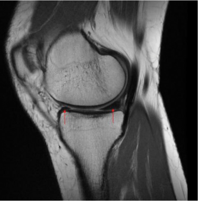
Context: Information regarding the structure, composition, and function of the knee menisci has been scattered across multiple sources and fields. This review contains a concise, detailed description of the knee menisci-including anatomy, etymology, phylogeny, ultrastructure and biochemistry, vascular anatomy and neuroanatomy, biomechanical function, maturation and aging, and imaging modalities.

Evidence acquisition: A literature search was performed by a review of PubMed and OVID articles published from 1858 to 2011.

Results: This study highlights the structural, compositional, and functional characteristics of the menisci, which may be relevant to clinical presentations, diagnosis, and surgical repairs.

Conclusions: An understanding of the normal anatomy and biomechanics of the menisci is a necessary prerequisite to understanding the pathogenesis of disorders involving the knee.


Figures

Figure 1.
Figure 1.
Gross photograph of human tibial plateau demonstrating the relative size and attachments of the medial and lateral menisci. The medial and lateral menisci (left side and right side of image, respectively) are connected by a transverse ligament (TL). 1, anterior insertional ligament of the medial meniscus; 2, posterior insertional ligament of the medial meniscus; 3, anterior insertional ligament of the lateral meniscus; 4, posterior insertional ligament of the lateral meniscus. ACL, anterior cruciate ligament; PCL, posterior cruciate ligament. Reprinted with permission from Messner and Gao.
Figure 2.
Figure 2.
(A) Anatomy of the meniscus viewed from above (adapted image reprinted with permission from Greis et al; original from Pagnani et al). (B) Axial view of a right tibial plateau showing sections of the meniscus and their relationship to the cruciate ligaments. AL, anterior horn lateral meniscus; AM, anterior horn medial meniscus; PCL, posterior cruciate ligament; PL, posterior horn lateral meniscus; PM, posterior horn medial meniscus. Reprinted with permission from Johnson et al.
Figure 3.
Figure 3.
Schematic diagram demonstrating the collagen fiber ultrastructure and orientation within the meniscus: 1, superficial network; 2, lamellar layer; 3, central main layer. Arrowheads, radial interwoven fibers; arrow, loose connective tissue. Reprinted with permission from Petersen and Tillmann.
Figure 4.
Figure 4.
Extracellular matrix. (A) Electron micrograph of an aggrecan aggregate shadowed by platinum. Many free aggrecan molecules are also seen. (B) Schematic drawing of an aggrecan aggregate shown in part A. Reprinted with permission from Alberts et al.
Figure 5.
Figure 5.
Confluence of geniculate arteries (anterior view). Reprinted with permission from Brindle et al.
Figure 6.
Figure 6.
Microvasculature of the medial meniscus (superior aspect), following vascular perfusion with India ink and tissue clearing using a modified Spälteholz technique. Perimeniscal capillary plexus (PCP) can be seen penetrating the peripheral border of the medial meniscus. F, femur; T, tibia. Reprinted with permission from Arnoczky and Warren.
Figure 7.
Figure 7.
Diagrams showing the mean movement (mm) in each meniscus during flexion (shaded) and extension (hashed). ANT, anterior; POST, posterior; mme, mean meniscal excursion; P/A, ratio of posterior to anterior meniscal translation during flexion. Reproduced with permission from Thomspon et al. *P < 0.05 (Student t test analysis).
Figure 8.
Figure 8.
Free body diagram of forces acting on the meniscus during loading. As the femur presses down on the meniscus during normal loading, the meniscus deforms radially but is anchored by its anterior and posterior horns (Fant and Fpost). During loading, tensile, compressive, and shear forces are generated. A tensile hoop stress (Fcir) results from the radial deformation, while vertical and horizontal forces (Fv and Fh) result from the femur pressing on the curved superior surface of the tissue. A radial reaction force (Frad) balances the femoral horizontal force (Fh). Reprinted with permission from Athanasiou and Sanchez-Adams.
Figure 9.
Figure 9.
A sagittal magnetic resonance (proton-density) image of a healthy knee depicting the medial menisci (arrows). The concave superior meniscal surface improves contact with the femoral epicondyles, and a flat undersurface improves contact with the tibial plateau. The periphery is thicker than the central portion, allowing for firm attachment to the joint capsule.

References

    1. Adams ME, Hukins DWL. The extracellular matrix of the meniscus. In: Mow VC, Arnoczky SP, Jackson DW. eds. Knee Meniscus: Basic and Clinical Foundations. New York, NY: Raven Press; 1992:15-282016
    1. Adams ME, McDevitt CA, Ho A, Muir H. Isolation and characterization of high-buoyant-density proteoglycans from semilunar menisci. J Bone Joint Surg Am. 1986;68:55-64 - PubMed
    1. Adams ME, Muir H. The glycosaminoglycans of canine menisci. Biochem J. 1981;197:385-389 - PMC - PubMed
    1. Ahmed AM, Burke DL. In-vitro measurement of static pressure distribution in synovial joints: part I. Tibial surface of the knee. J Biomech Eng. 1983;185:290-294 - PubMed
    1. Akgun U, Kogaoglu B, Orhan EK, Baslo MB, Karahan M. Possible reflex pathway between medial meniscus and semi-membranous muscle: an experimental study in rabbits. Knee Surg Sports Traumatol Arthrosc. 2008;16(9):809-814 - PubMed