. 2013 Jan 1;449(1):275-84.
doi: 10.1042/BJ20120625.

TfR1 interacts with the IKK complex and is involved in IKK-NF-κB signalling

Affiliations

TfR1 interacts with the IKK complex and is involved in IKK-NF-κB signalling

Niall S Kenneth et al. Biochem J. .

Abstract

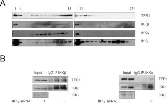
The IKK [inhibitor of NF-κB (nuclear factor κB) kinase] complex has an essential role in the activation of the family of NF-κB transcription factors in response to a variety of stimuli. To identify novel IKK-interacting proteins, we performed an unbiased proteomics screen where we identified TfR1 (transferrin receptor 1). TfR1 is required for transferrin binding and internalization and ultimately for iron homoeostasis. TfR1 depletion does not lead to changes in IKK subunit protein levels; however, it does reduce the formation of the IKK complex, and inhibits TNFα (tumour necrosis factor α)-induced NF-κB-dependent transcription. We find that, in the absence of TfR1, NF-κB does not translocate to the nucleus efficiently, and there is a reduction in the binding to target gene promoters and consequentially less target gene activation. Significantly, depletion of TfR1 results in an increase in apoptosis in response to TNFα treatment, which is rescued by elevating the levels of RelA/NF-κB. Taken together, these results indicate a new function for TfR1 in the control of IKK and NF-κB. Our data indicate that IKK-NF-κB responds to changes in iron within the cell.


Figures

Figure 1
Figure 1. TfR1 interacts with IKK in cells
(A) TfR1 interacts with TAP–IKKβ. TAP-tag-purified material was analysed by Western blotting for the presence of TfR1. (B) HEK-293 cells were transfected with the plasmids indicated and whole-cell lysates were prepared. Anti-GFP antibody-bound beads were used to immunoprecitate GFP–TfR1. Precipitates were resolved by SDS/PAGE and then analysed by Western blotting using the antibodies indicated. (C) Whole-cell lysates were prepared from U2OS cells and immunoprecipitated with normal rabbit IgG or anti-IKK antibodies. Precipitates were resolved by SDS/PAGE and then analysed by Western blotting using the antibodies indicated. (D) U20S cells were treated with 10 ng/ml TNFα for the times indicated before lysis. Whole-cell lysates were immunoprecipitated with normal rabbit IgG or anti-IKK antibodies. Precipitates were processed and analysed as in (C). IP, immunoprecipitation.
Figure 2
Figure 2. TfR1 forms several complexes with the IKK proteins
(A) Whole-cell lysates prepared from U2OS cells were run on a size-exclusion chromatography column, and gradient-eluted fractions (1–26) were immunoblotted with the antibodies indicated. (B) U2OS cells were transfected with control and IKKγ siRNA oligonucleotides before lysis. Immunoprecipitations were performed for IKKβ and IKKα, with IgG as a control. Precipitates were resolved by SDS/PAGE and then analysed by Western blotting using the antibodies indicated. IP, immunoprecipitation.
Figure 3
Figure 3. Depletion of TfR1 impairs IKK complex formation
(A) U2OS cells were transiently depleted of TfR1 using siRNA. Whole-cell lysates were subjected to immunoblot analysis for the levels of the proteins indicated. (B) Whole-cell lysates prepared from U2OS cells treated with non-targeting (NT) siRNA or TfR1 siRNAs were immunoprecipitated with normal rabbit IgG or anti-IKKγ or anti-IKKβ antibodies. Precipitates were resolved by SDS/PAGE and then analysed by Western blotting with anti-IKKα, anti-IKKβ and anti-IKKγ antibodies to assess the integrity of the IKK complex. IP, immunoprecipitation.
Figure 4
Figure 4. Depletion of TfR1 or iron impairs IKK and NF-κB activity
(A) U2OS cells were depleted of TfR1 using siRNA and subsequently treated with 10 ng/ml TNFα for the times indicated. Whole-cell lysates were subjected to immunoblot analysis for the levels of the proteins indicated. (B) U2OS-NF-κB luciferase reporter cells were transfected with the siRNAs indicated and treated with 10 ng/ml TNFα for 5 h before harvest. Results are means±S.E.M. for at least three independent experiments expressed as fold activation/repression relative to TNFα-treated control siRNA levels. (C) U2OS-NF-κB luciferase reporter cells were transfected with 1 μg of control or TfR1 expression constructs before treatment with 10 ng/ml TNFα for 5 h before harvest. Results are means±S.E.M. for at least three independent experiments expressed as fold activation/repression relative to controls. (D) U2OS-NF-κB reporter luciferase reporter cells were treated with 200 μM DFX or 300 μM iron/2 μM ascorbate 2 h before treatment with 10 ng/ml TNFα for an additional 5 h. Results are means±S.E.M. for at least three independent experiments expressed as fold activation/repression relative to control. (E) U2OS cells were treated with either DFX or iron as in (D), but TNFα was added for 24 h before lysis. Whole-cell lysates were subjected to immunoblot analysis for the levels of the proteins indicated. *P<0.05; **P<0.01; ***P<0.001.
Figure 5
Figure 5. Depletion of TfR1 impairs activation of endogenous NF-κB-target genes
(A) Nuclear and cytoplasmic extracts were prepared from control cells or cells depleted of TfR1, and subsequently treated with TNFα for the times indicated. Nuclear extracts were analysed by Western blotting using the antibodies indicated. Whole-cell lysates were analysed for total levels of RelA under the same conditions. Levels of nuclear RelA were quantified and are shown below the RelA panel on the left. (B) Quantitative RT–PCR analysis of p100 and c-IAP2 mRNA prepared from U2OS cells depleted of TfR1 and subsequently stimulated with TNFα for the times indicated. ***P<0.01. (C) U2OS cells were depleted of TfR1, and subsequently stimulated with TNFα for the times indicated. Whole-cell lysates were subjected to immunoblot analysis for the levels of the proteins indicated. (D) ChIP assay showing cross-linking of RelA to the NF-κB consensus sequence and a control region in the p100 promoter, in cells transfected with non-targeting siRNA or cells transfected with siRNA against TfR1, in the presence or absence of 10 ng/ml TNFα stimulation for 1 h. Ab, antibody; NT, targeting.
Figure 6
Figure 6. Depletion of TfR1 sensitizes cells to TNFα-induced apoptosis
(A) U2OS cells were depleted of TfR1, and subsequently stimulated with increasing concentrations (0, 10, 20, 30 and 40 ng/ml) of TNFα for 24 h. Whole-cell lysates were subjected to immunoblotting analysis for the levels of the proteins indicated. NT, non-targeting. (B) U2OS cells were transfected with 1 μg of control or RelA-expression constructs 24 h before TfR1 depletion by siRNA. Cells were treated with 10 ng/ml TNFα for an additional 24 h before lysis. Whole-cell lysates were subjected to immunoblotting analysis for the levels of the proteins indicated.

Similar articles

Cited by

References

    1. Perkins N. D. The diverse and complex roles of NF-κB subunits in cancer. Nat. Rev. Cancer. 2012;12:121–132. - PubMed
    1. Perkins N. D. Integrating cell-signalling pathways with NF-κB and IKK function. Nat. Rev. Mol. Cell Biol. 2007;8:49–62. - PubMed
    1. Israel A. The IKK complex, a central regulator of NF-κB activation. Cold Spring Harbor Perspect. Biol. 2010;2:a000158. - PMC - PubMed
    1. Wertz I. E., Dixit V. M. Signaling to NF-κB: regulation by ubiquitination. Cold Spring Harbor Perspect. Biol. 2010;2:a003350. - PMC - PubMed
    1. Comb W. C., Cogswell P., Sitcheran R., Baldwin A. S. IKK-dependent, NF-κB-independent control of autophagic gene expression. Oncogene. 2011;30:1727–1732. - PMC - PubMed

Publication types