. 2012 May-Jun;14(3):277-85.
doi: 10.1111/j.1525-142X.2012.00544.x.

Evolution of electrosensory ampullary organs: conservation of Eya4 expression during lateral line development in jawed vertebrates

Affiliations

Evolution of electrosensory ampullary organs: conservation of Eya4 expression during lateral line development in jawed vertebrates

Melinda S Modrell et al. Evol Dev. 2012 May-Jun.

Abstract

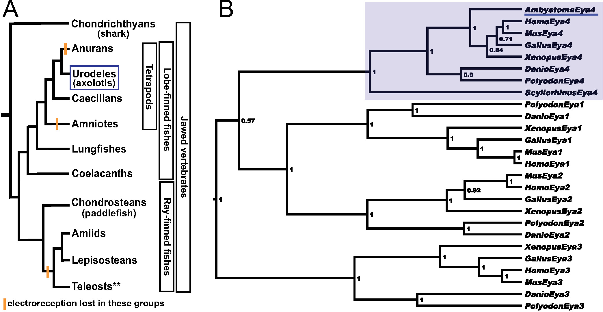
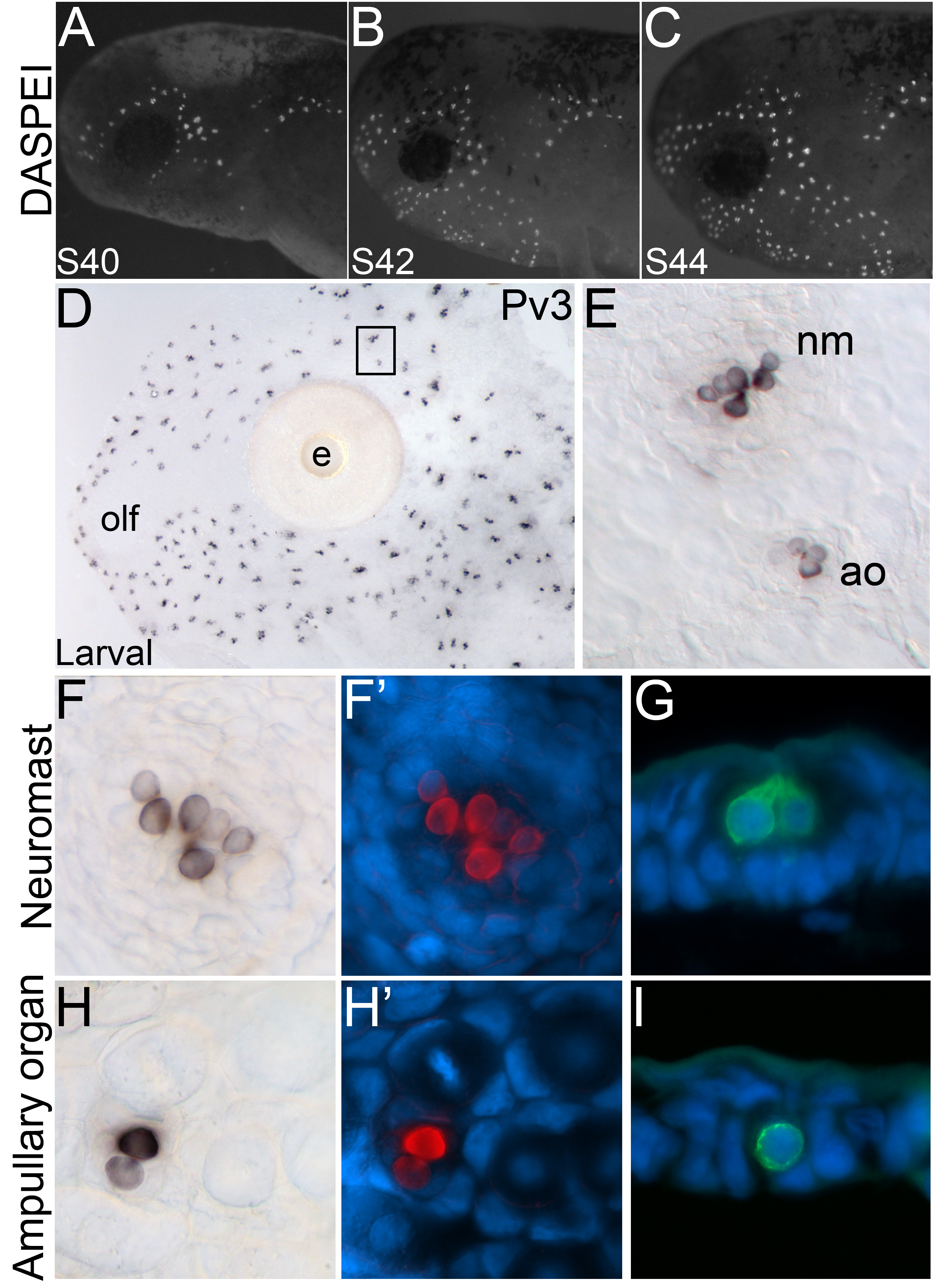
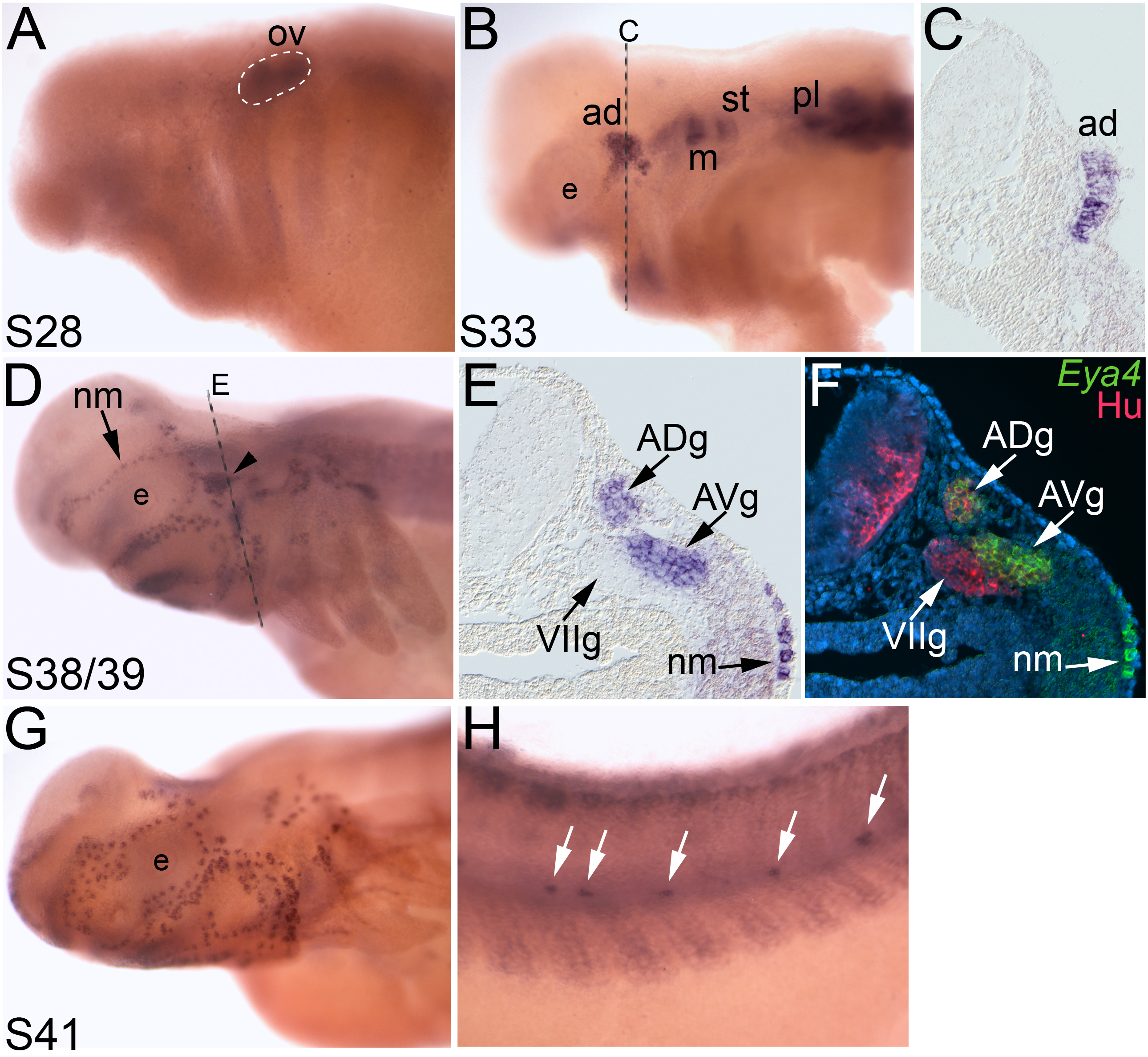
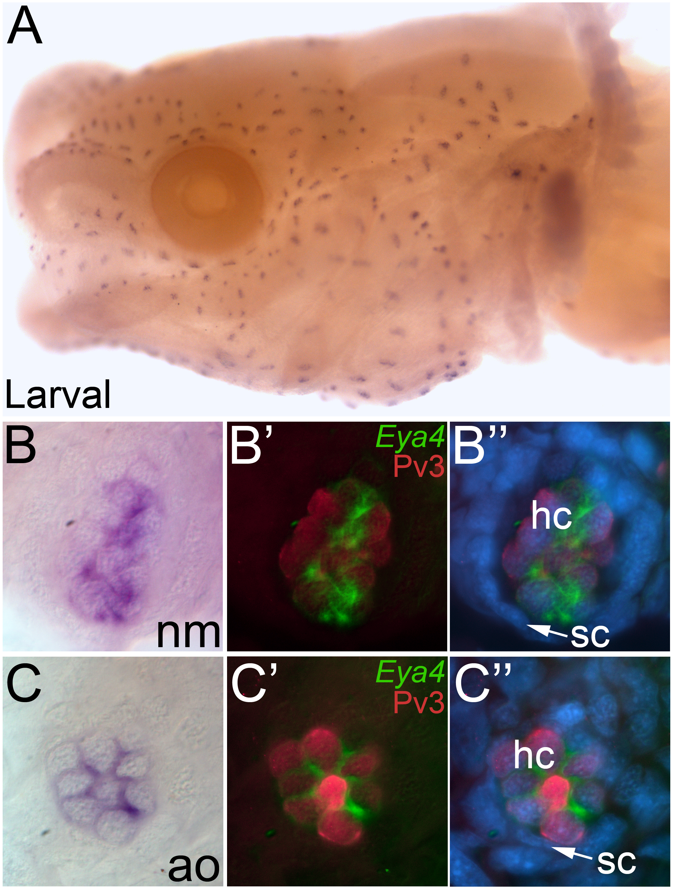
The lateral line system of fishes and amphibians comprises two ancient sensory systems: mechanoreception and electroreception. Electroreception is found in all major vertebrate groups (i.e. jawless fishes, cartilaginous fishes, and bony fishes); however, it was lost in several groups including anuran amphibians (frogs) and amniotes (reptiles, birds, and mammals), as well as in the lineage leading to the neopterygian clade of bony fishes (bowfins, gars, and teleosts). Electroreception is mediated by modified "hair cells," which are collected in ampullary organs that flank lines of mechanosensory hair cell containing neuromasts. In the axolotl (a urodele amphibian), grafting and ablation studies have shown a lateral line placode origin for both mechanosensory neuromasts and electrosensory ampullary organs (and the neurons that innervate them). However, little is known at the molecular level about the development of the amphibian lateral line system in general and electrosensory ampullary organs in particular. Previously, we identified Eya4 as a marker for lateral line (and otic) placodes, neuromasts, and ampullary organs in a shark (a cartilaginous fish) and a paddlefish (a basal ray-finned fish). Here, we show that Eya4 is similarly expressed during otic and lateral line placode development in the axolotl (a representative of the lobe-finned fish clade). Furthermore, Eya4 expression is specifically restricted to hair cells in both neuromasts and ampullary organs, as identified by coexpression with the calcium-buffering protein Parvalbumin3. As well as identifying new molecular markers for amphibian mechanosensory and electrosensory hair cells, these data demonstrate that Eya4 is a conserved marker for lateral line placodes and their derivatives in all jawed vertebrates.


Figures

Fig. 1
Fig. 1. Vertebrate phylogeny showing the distribution of electroreception and the molecular phylogenetic analysis of Eya4..
(A) Simplified phylogeny of the jawed vertebrates (gnathostomes). Electroreception is found in all major groups including both major clades of bony fishes, the lobe-finned fishes and the ray-finned fishes, but was lost in lineages indicated by orange bars. **Within teleosts, several groups independently evolved electroreception. (B) Phylogenetic tree showing the orthology of the Ambystoma mexicanum Eya4 cDNA fragment with other vertebrate Eya4 homologues (Eya4 clade is shaded in light blue). A. mexicanum Eya4 is underlined in blue. Numbers at nodes represent the Bayesian posterior probability of each clade. Species abbreviations are as follows: Ambystoma, Ambystoma mexicanum (axolotl); Danio, Danio rerio (zebrafish); Gallus, Gallus gallus (chicken); Homo, Homo sapiens (human); Mus, Mus musculus (mouse); Polyodon, Polyodon spathula (North American paddlefish); Scyliorhinus, Scyliorhinus canicula (lesser-spotted dogfish/catshark); Xenopus, Xenopus tropicalis (Western/tropical clawed frog).
Fig. 2
Fig. 2. Spatiotemporal development of sensory hair cells of axolotl lateral line organs.
Anterior to the left; lateral views. (A-C) Fluorescent images superimposed on darkfield images of stage 40 through 44 embryos stained with the vital dye DASPEI (white), which selectively labels functional neuromasts after a short incubation. (A) Stage 40 embryo. Neuromasts derived from the anteriormost lateral line placodes are already present. (B) Stage 42 embryo. (C) Stage 44 embryo. (D-I) (D) Skinmount from a larva (1-2 cm in length) immunostained for Pv3, showing expression in mature lateral line organs. (E) Higher power view of the boxed area in A. Hair cells in both the mechanosensory neuromasts and electrosensory ampullary organs are labelled by Pv3. (F) Surface view of a single neuromast showing Pv3-positive hair cells (black) in a brightfield image and (F’) in a false color overlay (Pv3, red) with the nuclear marker DAPI (blue). In this view, approximately seven individual hair cells can be identified. (G) Transverse section through a neuromast showing Pv3-positive hair cells (green) and DAPI (blue). (H) Surface view of a single ampullary organ showing Pv3-positive hair cells (black) in a brightfield image and (H’) in a false color overlay (Pv3, red) with DAPI (blue). Ampullary organs contain fewer hair cells per organ than neuromasts: in this example, there are only two hair cells. (I) Transverse section through an ampullary organ showing a single Pv3-positive hair cell (green) with DAPI (blue). Abbreviations: ao, ampullary organ; e, eye; nm, neuromast; olf, olfactory organ.
Fig. 3
Fig. 3. Eya4 expression in lateral line placodes and ganglia.
In situ hybridizations of whole-mount axolotl embryos showing the localization of Eya4. Anterior to the left; lateral views unless otherwise noted. (A) Stage 28 embryo. Eya4 expression is restricted to the otic vesicle (outlined) and weakly in the developing somites. (B) By stage 33, several lateral line placodes have begun to express Eya4. (C) Transverse section through the anterodorsal lateral line placode (ad) indicated in B by the dashed line. Eya4 expression is restricted to the thickened epithelium (i.e. the placode). (D) At stages 38-39, Eya4 expression is observed in neuromasts (arrow) and lateral line ganglia (arrowhead). Dashed line indicates plane of section shown in E. (E) Transverse section through the region indicated in D by the dashed line. Eya4 is expressed in the anterodorsal and anteroventral lateral line ganglia, and in developing neuromasts beneath the surface ectoderm. (F) The same transverse section shown in E, immunostained for the panneuronal marker HuC/D (pink), with a false color overlay of the brightfield image in E, showing Eya4 (green) and the nuclear marker DAPI (blue). Eya4 is restricted to the lateral line ganglia and is not observed in other cranial ganglia, such as the ganglion of the facial (VII) nerve. (G) Stage 41 embryo. Neuromast expression of Eya4 continues as more organs differentiate. (H). Trunk of the stage 41 embryo shown in G. The posterior trunk neuromasts also express Eya4 (arrows). Abbreviations: ad, anterodorsal lateral line placode; ADg, ganglion of the anterodorsal lateral line nerve; AVg, ganglion of the anteroventral lateral line nerve; e, eye; m, middle lateral line placode; nm, neuromast; ov, otic vesicle; pl, posterior lateral line placode; st, supratemporal lateral line placode; VIIg, ganglion of the facial (VII) nerve.
Fig. 4
Fig. 4. Eya4 is restricted to the hair cells in mature neuromasts and ampullary organs.
Whole-mount in situ hybridization for Eya4 in larval axolotls. (A) Head view of a larval axolotl. Anterior to the left, lateral view. Eya4 expression is observed in both the neuromasts and ampullary organs. (B) Higher power view of a single neuromast, showing the restricted localization of Eya4. (B’, B”) Fluorescent image of the neuromast in B, immunostained for the hair cell marker Pv3 (red) with Eya4 as a false colored overlay (green) plus (in B”) the nuclear marker DAPI (blue). (C) Higher power view of a single ampullary organ showing the restricted localization of Eya4. (C’, C”) Fluorescent image of the ampullary organ in C, immunostained for the hair cell marker Pv3 (red) with Eya4 as a false colored overlay (green), plus (in C”) the nuclear marker DAPI (blue). In both neuromasts and ampullary organs, Eya4 transcripts are localized within the cytoplasm of the Pv3-positive hair cells. Abbreviations: ao, ampullary organ; hc, hair cell; nm, neuromast; sc, support cell.

References

    1. Alves-Gomes JA. The evolution of electroreception and bioelectrogenesis in teleost fish: a phylogenetic perspective. J. Fish Biol. 2001;58:1489–1511.
    1. Balak KJ, Corwin JT, Jones JE. Regenerated hair cells can originate from supporting cell progeny: evidence from phototoxicity and laser ablation experiments in the lateral line system. J. Neurosci. 1990;10:2502–2512. - PMC - PubMed
    1. Bessarab DA, Chong SW, Korzh V. Expression of zebrafish six1 during sensory organ development and myogenesis. Dev. Dyn. 2004;230:781–786. - PubMed
    1. Bodznick D, Montgomery JC. The physiology of low-frequency electrosensory systems. In: Bullock TH, Hopkins CD, Popper AN, Fay RR, editors. Electroreception. Springer; New York: 2005. pp. 132–153.
    1. Bullock TH, Hopkins CD, Popper AN, Fay RR, editors. Electroreception. Springer; New York: 2005.

MeSH terms