Review
. 2013 Jun;54(2):250-7.
doi: 10.1016/j.bone.2012.09.016. Epub 2012 Sep 24.

Effects of PTH on osteocyte function

Affiliations
Review

Effects of PTH on osteocyte function

Teresita Bellido et al. Bone. 2013 Jun.

Abstract

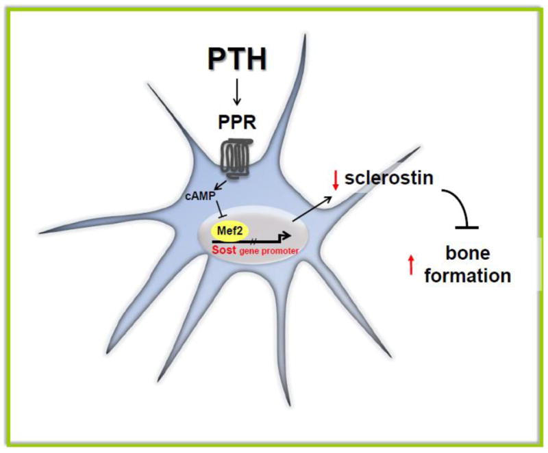
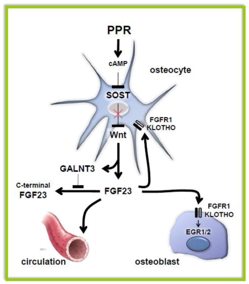
Osteocytes are ideally positioned to detect and respond to mechanical and hormonal stimuli and to coordinate the function of osteoblasts and osteoclasts. However, evidence supporting the involvement of osteocytes in specific aspects of skeletal biology has been limited mainly due to the lack of suitable experimental approaches. Few crucial advances in the field in the past several years have markedly increased our understanding of the function of osteocytes. The development of osteocytic cell lines initiated a plethora of in vitro studies that have provided insights into the unique biology of osteocytes and continue to generate novel hypotheses. Genetic approaches using promoter fragments that direct gene expression to osteocytes allowed the generation of mice with gain or loss of function of particular genes revealing their role in osteocyte function. Furthermore, evidence that Sost/sclerostin is expressed primarily in osteocytes and inhibits bone formation by osteoblasts, fueled research attempting to identify regulators of this gene as well as other osteocyte products that impact the function of osteoblasts and osteoclasts. The discovery that parathyroid hormone (PTH), a central regulator of bone homeostasis, inhibits sclerostin expression generated a cascade of studies that revealed that osteocytes are crucial target cells of the actions of PTH. This review highlights these investigations and discusses their significance for advancing our understanding of the mechanisms by which osteocytes regulate bone homeostasis and for developing therapies for bone diseases targeting osteocytes.


Figures

Figure 1
Figure 1. PTH downregulates Sost/sclerostin expression in osteocytes
PPR activation by PTH elevates cAMP levels and inhibits Mef2-stimulated Sost promoter activity leading to decreased expression of the inhibitor of bone formation sclerostin, and elevated bone formation rate.
Figure 2
Figure 2. Converse effects of PPR activation and PPR deletion in osteocytes on bone formation and resorption
Osteocyte activation of the PPR leads to cAMP-dependent Sost downregulation and increased Wnt signaling, which in turn increases osteoblasts and stimulates bone formation. PPR activation increases RANKL expression, osteoclasts and resorption. However, OPG expression is not affected, likely resulting from opposing effects on the expression of the gene by cAMP (reduction) and Wnts (elevation). The main effect of deletion of the PPR in osteocytes is increased SOST expression, decreased RANKL expression, reduced osteoclast and osteoblast number with decreased bone resorption and bone formation, depending on the cKO model used (see text for details).
Figure 3
Figure 3. PPR signaling modulates the endocrine and auto/paracrine function of osteocytes by regulating FGF23 expression
cAMP elevation in vivo by a constitutively active PPR or in vitro by the PPR ligands PTH and PTHrP increases FGF23 in a Wnt-dependent manner. A simultaneous increase in GALNT3 stabilizes FGF23, leading to increased intact FGF23 in the circulation as well as locally in bone. Binding of FGF23 to the FGFR1/KLOTHO receptor complex expressed in osteocytes and osteoblasts activates intracellular signaling.
Figure 4
Figure 4. PTH receptor and osteocytic lacunae/canalicular remodeling during lactation
During lactation, increased circulating levels of PTHrP activate PPR expressed in osteocytes and induce increase in lacuna/canalicular remodeling and consequent bone loss. Genes such as TRAP, Cathepsin K and carbonic anydrase 2 are highly expressed in osteocytes from lactating mice and their regulation is dependent on PPR expression.

References

    1. Aarden EM, Burger EH, Nijweide PJ. Function of osteocytes in bone. J Cell Biochem. 1994;55:287–99. - PubMed
    1. Bonewald LF. The Amazing Osteocyte. J Bone Miner Res. 2011;26:229–38. - PMC - PubMed
    1. Winkler DG, Sutherland MK, Geoghegan JC, Yu C, Hayes T, Skonier JE, Shpektor D, Jonas M, Kovacevich BR, Staehling-Hampton K, Appleby M, Brunkow ME, Latham JA. Osteocyte control of bone formation via sclerostin, a novel BMP antagonist. EMBO J. 2003;22:6267–76. - PMC - PubMed
    1. Van Bezooijen RL, Roelen BA, Visser A, Wee-Pals L, de Wilt E, Karperien M, Hamersma H, Papapoulos SE, Ten Dijke P, Lowik CW. Sclerostin ss an osteocyte-expressed negative regulator of bone formation, but not a classical BMP antagonist. J Exp Med. 2004;199:805–14. - PMC - PubMed
    1. Bellido T, Ali AA, Gubrij I, Plotkin LI, Fu Q, O’Brien CA, Manolagas SC, Jilka RL. Chronic elevation of PTH in mice reduces expression of sclerostin by osteocytes: a novel mechanism for hormonal control of osteoblastogenesis. Endocrinology. 2005;146:4577–83. - PubMed

Substances