. 2012 Oct;44(10):876-85.
doi: 10.1093/abbs/gms072.

Cloning and primary characterizations of rLcn9, a new member of epididymal lipocalins in rat

Affiliations

Cloning and primary characterizations of rLcn9, a new member of epididymal lipocalins in rat

Xiangqi Li et al. Acta Biochim Biophys Sin (Shanghai). 2012 Oct.

Abstract

Lipocalins are a structurally conserved and diversely functional family of proteins that are of potential importance in epididymis functions. The rat Lcn9 gene was cloned by in silico methods and genome walking based on homology to the rhesus monkey epididymal ESC513 and its polyclonal antisera were prepared. The rat Lcn9 gene is located on chromosome 3p13 spanning 7 exons, contains 2.3 kb and encodes 179 amino acids with a 17-amino acid signal peptide. Northern blot, western blot, and immunohistochemical staining analysis revealed that rat Lcn9 was a novel epididymis-specific gene, expressed selectively in the proximal caput region, influenced by luminal fluid testicular factors. Moreover, Lcn9 protein was modified by N-glycosylation and bound on the postacrosomal domain of caput sperm. In conclusion, the rat Lcn9 exhibited tissue-, region-, and temporal-specific expression patterns and its expression was regulated by luminal testicular factors. Its potential roles in sperm maturation are discussed.


Figures
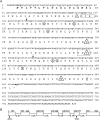
Figure 1
Figure 1
Rat Lcn9 gene structure and cDNA and protein sequence (A) The rLcn9 open reading frame (ORF) contains 540 bp coding for a 179-amino-acid protein, and the initial and terminal codons are indicated in bold. The protein contains conserved motifs (GVW, TDY, R, and C) indicated by ‘□’; a putative signal peptide (italics and bold) with a cleavage site between amino acids 17 and 18; three N-glycosylation sites (N47, N68, N130) indicated by ‘○’; three serine, threonine, and tyrosine phosphorylation sites (S35, S97, T172, Y117) indicated by ‘▵’. Two ubiquitination sites (K129 and K54) indicated by ‘◊’. Sequences between two arrows (134–378 bp) were used as the probe for the northern blot analysis. (B) The rat LCN9 gene contains 7 exons represented by open boxes. The nucleotides in the cDNA are indicated by the numbers above the boxes. The numbers between the boxes indicate the sizes of the introns.
Figure 2
Figure 2
Amino acid sequences alignment of rat lipocalin genes (Lcn2, Lcn5, Lcn6, and Lcn8–Lcn13) with mouse Lcn9 Dashes show alignment gaps. The conserved motifs (G-X-W, T-D-Y, R, and C) of the lipocalin family are indicated by red color. The putative signal peptides are indicated by blue color. Amino acid residue numbers are shown above and on the left.
Figure 3
Figure 3
Selective expression of Lcn9 mRNA in the rat epididymis Northern blot analysis shows the cn9 mRNA is highly expressed in the caput region, but not in the other regions or tissues. Ca, caput; Co, corpus; Cu, cauda; Te, testis; Vp, ventral prostate; Sp, spleen; He, heart; Br, brain; Si, small intestine; Lu, lung; Li, liver; St, stomach; Ki, kidney; Sv, seminal vesicle; Pa, pancreas; Th, thyroid; Vd, vas deference.
Figure 4
Figure 4
Lcn9 expression after androgen ablation (A) Northern blot analysis. 0d was prior to castration; 1d, 3d, 5d, and 7d indicated days after bilateral castration; and 7d+1, 7d+3, 7d+5, and 7d+7 indicated days after the initial injection of testosterone propionate applied to the 7d-castrated rats. Injections were continued every 2 days. (B) Comparison of Lcn9 mRNA and the serum testosterone levels. Left Y axis is hybridization density of Lcn9 mRNA/18S ribosomal RNA in the rat epididymis, while right Y axis is the serum testosterone level (expressed in nM). The RNAs were pooled from four to seven animals per group.
Figure 5
Figure 5
Western blot analysis of rat rLcn9 recombinant protein and native protein (A) The sensitivity analysis of rLcn9 antibody with 0.5, 1, and 2 ng of the antigen peptide. (B) The specificity analysis of rLcn9 antibody with rat native protein. Ca, caput epididymidis; Co, corpus epididymidis; Cu, cauda epididymidis; Te, testis; Ep, epididymis; Vd, vas deferens; Pr, prostate; Ki, kidney; He, heart; Li, liver; Lu, lung; Sp, spleen; In, intestine; Br, brain. (C) The change of molecular masses of rat rLcn9 before and after deglycosylation by PNGase-F.
Figure 6
Figure 6
The localization of rLcn9 in the 120-day-old rat epididymis by immunohistochemistry (A) The rat rLcn9 localization in different regions of the epididymis. The photograph is from the scanning of the section by Magicscan V4.6. (B) The expression pattern of rLcn9 in the whole rat epididymis. The magnified photograph of the caput region in (A). (C) The magnified photographs of some parts of (A): 1–9, caput; 10, proximal corpus; 11, distal corpus; 12, cauda. Magnification, ×40. (Note: parts here are corresponding to segments reported before [38]: part 1 to segment 1, parts 2 and 3 to segment 2, parts 4 and 5 to segment 3, part 6 to segment 4.)
Figure 7
Figure 7
The rat LCN9 localization in different regions of the rat epididymis sections from different developmental stages by immunohistochemistry The ages (expressed in days) are shown on the left, and the regions of the epididymis are shown at the top.
Figure 8
Figure 8
Lcn9 localization on the caput spermatozoa by immunofluorescence (A) Immunolocalization of rat Lcn9 on spermatozoa isolated from the caput epididymal region by pre-immune sera. (B) Immunolocalization of rat Lcn9 on spermatozoa isolated from the caput epididymal region by rLcn9 antibodies. (C) Magnified heads of spermatozoa in (B). Magnification, ×100. (a1, b1, c1) Phase contrast view of sperm in a2, b2, c2; (a2, b2, c2) The immunofluorescence of rat rLcn9 (FITC labeled); (a3, b3, c3) The nuclear (PI); (a4, b4, c4) The merged photograph of a1 and a2, b1 and b2, and c1 and c2, respectively; (a5, b5, c5) The merged photograph of a3 and a4, b3 and b4, and c3 and c4, respectively.

References

    1. Banks S, King SA, Irvine DS, Saunders PT. Saunders. Impact of a mild scrotal heat stress on DNA integrity in murine spermatozoa. Reproduction. 2005;129:505–514. - PubMed
    1. Dacheux JL. Protein secretion in the epididymis. In: Robaire B, Hinton BT, editors. The Epididymis: From Molecules to Clinical Practice. New York: Kluwer Academic/Plenum Publishers; 2002. pp. 151–168.
    1. Jones R. Plasma membrane composition and organisation during maturation of spermatozoa in the epididymis. In: Robaire B, Hinton BT, editors. The Epididymis: from Molecules to Clinical Practice. New York: Kluwer Academic/Plenum Publishers; 2002. pp. 405–416.
    1. Dacheux JL, Gatti JL, Dacheux F. Contribution of epididymal secretory proteins for spermatozoa maturation. Microsc Res Tech. 2003;61:7–17. - PubMed
    1. Khole V. Epididymis as a target for contraception. Indian J Exp Biol. 2003;41:764–772. - PubMed

Publication types

MeSH terms

Associated data