. 2012 Nov 1;189(9):4496-509.
doi: 10.4049/jimmunol.1201121. Epub 2012 Sep 26.

Targeting the genital tract mucosa with a lipopeptide/recombinant adenovirus prime/boost vaccine induces potent and long-lasting CD8+ T cell immunity against herpes: importance of MyD88

Affiliations

Targeting the genital tract mucosa with a lipopeptide/recombinant adenovirus prime/boost vaccine induces potent and long-lasting CD8+ T cell immunity against herpes: importance of MyD88

Xiuli Zhang et al. J Immunol. .

Abstract

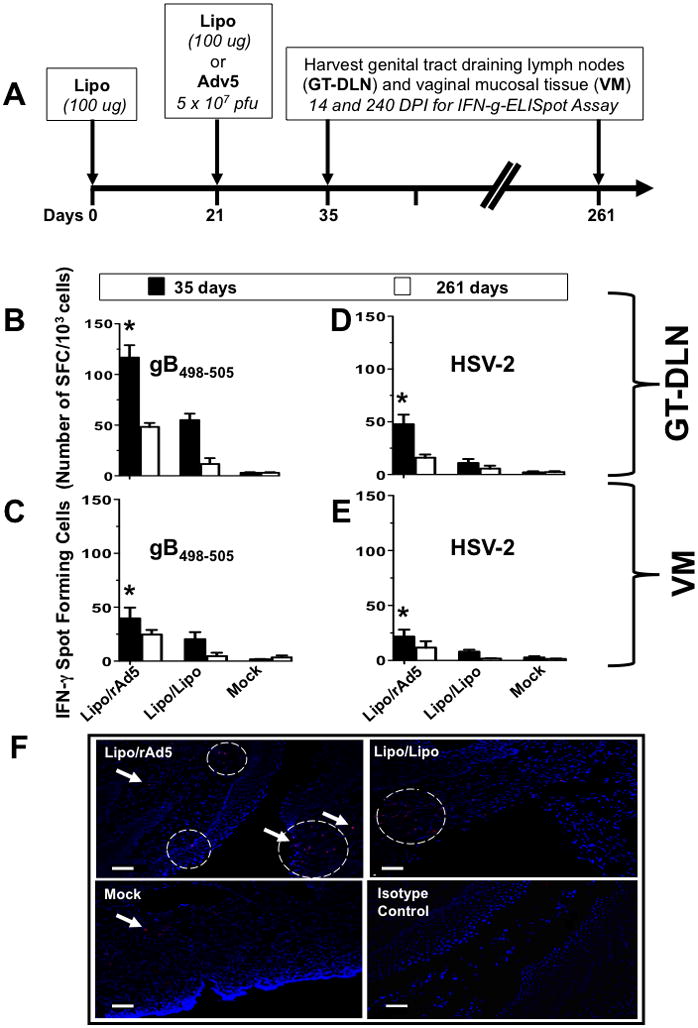
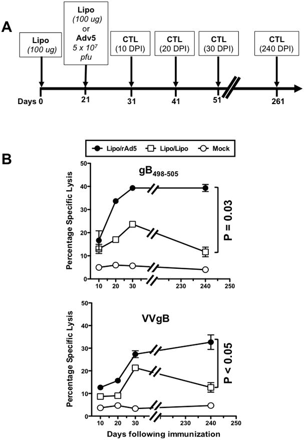
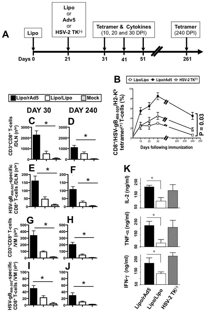
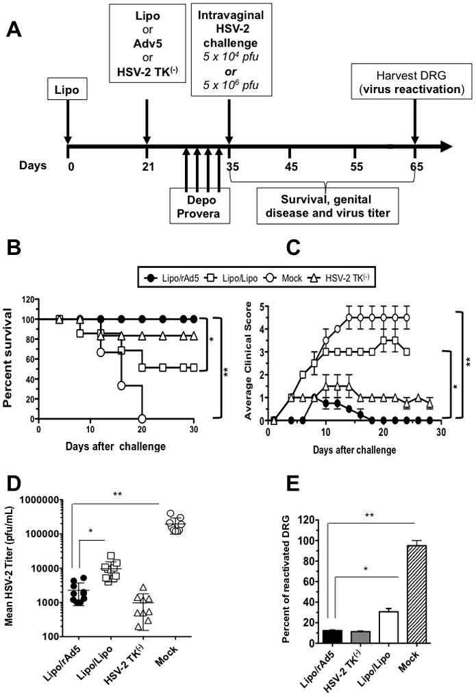
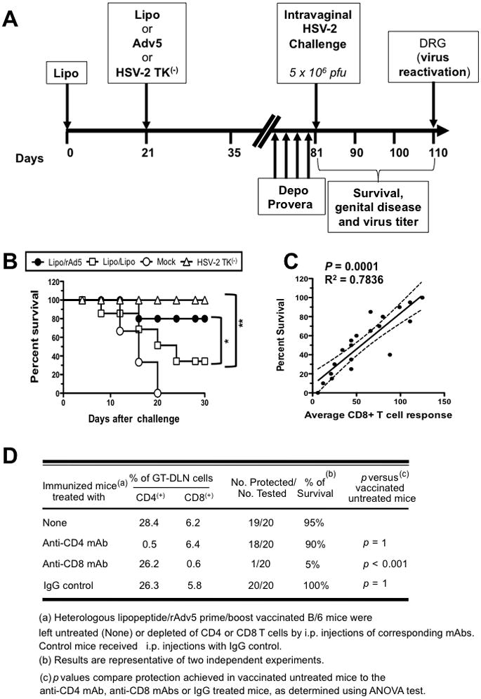
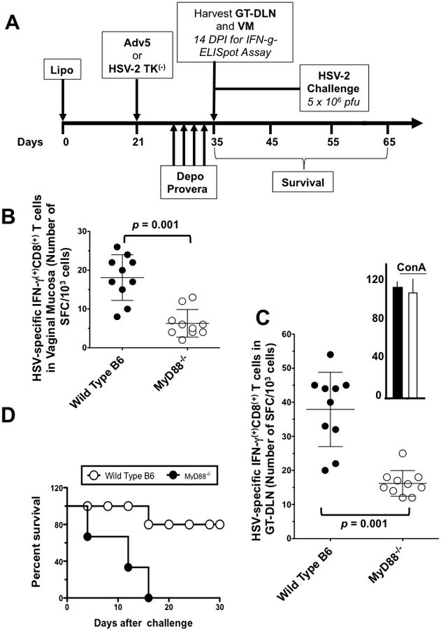
Targeting of the mucosal immune system of the genital tract with subunit vaccines has failed to induce potent and durable local CD8(+) T cell immunity, which is crucial for protection against many sexually transmitted viral pathogens, including HSV type 2 (HSV-2), which causes genital herpes. In this study, we aimed to investigate the potential of a novel lipopeptide/adenovirus type 5 (Lipo/rAdv5) prime/boost mucosal vaccine for induction of CD8(+) T cell immunity to protect the female genital tract from herpes. The lipopeptide vaccine and the rAdv5 vaccine express the immunodominant HSV-2 CD8(+) T cell epitope (gB(498-505)), and both were delivered intravaginally in the progesterone-induced B6 mouse model of genital herpes. Compared with mice immunized with the homologous lipopeptide/lipopeptide (Lipo/Lipo) vaccine, the Lipo/rAdv5 prime/boost immunized mice 1) developed potent and sustained HSV-specific CD8(+) T cells, detected in both the genital tract draining nodes and in the vaginal mucosa; 2) had significantly lower virus titers; 3) had decreased overt signs of genital herpes disease; and 4) did not succumb to lethal infection (p < 0.005) after intravaginal HSV-2 challenge. Polyfunctional CD8(+) T cells, producing IFN-γ, TNF-α, and IL-2 and exhibiting cytotoxic activity, were associated with protection (p < 0.005). The protective CD8(+) T cell response was significantly compromised in the absence of the adapter MyD88 (p = 0.0001). Taken together, these findings indicate that targeting of the vaginal mucosa with a Lipo/rAdv5 prime/boost vaccine elicits a potent, MyD88-dependent, and long-lasting mucosal CD8(+) T cell protective immunity against sexually transmitted herpes infection and disease.


Figures

Figure 1
Figure 1. Potent and long-lasting CD8+ T cell responses detected in both GT-DLN and VM following IVAG immunization with Lipo/rAdv5 prime/boost vaccine
(A) Time course for immunization and CD8+ T cell response analysis. Four groups of B6 mice (n =10) were immunized IVAG with: 100 μg of lipopeptide in saline on days 0, and 21 (Lipo/Lipo), with lipopeptide on day 0 and then with 5 × 10⁷ pfu of rAd5 in saline on days 21 (Lipo/rAdv5 prime/boost mucosal vaccine or Lipo/rAd5), or with the irrelevant OVA257-264 lipopeptide and an empty Ad5 vector in saline on days 0 and 21 (mock-immunized or Mock). (BE) On day 35 and 261 (i.e. fourteen and 240 days post-immunization (DPI)), the iliac and inguinal lymph nodes draining the genital tract (GT-DLN) (B and D) as well as the vaginal mucosa (VM) (C and E) were harvested. GT-DLN and VM cell suspensions were assayed for gB498-505- (B and C) and HSV-2- (D and E) specific IFN-γ-producing CD8+ T cell responses using ELISpot assay, as described in Material & Methods. Values represent the mean of IFN-γ spot forming cells detected in an average of 5 mice. (*) Indicates the P values were ≤ 0.05 when HSV-2 or gB498-505-specific IFN-γ-secreting CD8+ T cell responses from the Lipo/rAdv5 vaccine group of mice was compared to the homologous Lipo/Lipo group or to the mock-immunized group (one-way ANOVA). (F) Vaginal mucosal tissue was collected 14 days after the second immunization from the progesterone-treated mice that were immunized with Lipo/rAdv5 (upper left), Lipo/Lipo (upper right) or mock-immunized (lower left) and CD8+ T cells were detected by immunofluorescence microscopy, as described in Material & Methods. Sections were stained with fluorescein isothiocyanate-conjugated anti-CD8 antibody, and the nuclei were visualized by staining with DAPI (blue). Arrowheads and circles indicate CD8+ T cells accumulating at the vaginal epithelium and stroma. Staining with an isotype control IgG is shown in the lower right picture. Scale bars = 50 υm. Results are representative of two independent experiments.
Figure 2
Figure 2. Kenetics of gB498-505-specific CTL responses induced by the Lipo/rAdv5 prime/boost vs. Lipo/Lipo mucosal vaccines
(A) Groups of B6 mice (n =40) were immunized IVAG with the Lipo/rAdv5 prime/boost mucosal vaccine in saline, or with the homologous Lipo/Lipo, or with the irrelevant OVA257-264 lipopeptide and empty Ad5 vector in PBS on days 0 and 21 (mock-immunized or Mock), similar to Fig. 1 above. On day 10, 20, 30 and 240 post-immunization 5 mice were euthanized per each time point/group, and CTL activity was assessed of GT-DLN derived effector CD8+ T-cells was determined in a CRA assay using as target cells autologous EL-4 (at E:T of 30) loaded with HSV-2 gB498-505 peptide (Fig. 2B upper panel) or transfected with VVgB, a vaccinia virus expressing gB (Fig. 2B lower panel). The data are representative of two independent experiments and the bars represent SD between the experiments. The p values show significance levels of differences in the overall amount of cytotoxic activity between Lipo/rAdv5 and Lipo/Lipo immunized mice (one way, ANOVA test).
Figure 3
Figure 3. Higher percentages and increased numbers of HSV-gB498-505-specific CD8+ T-cells induced by the Lipo/rAdv5 prime/boost compared to its Lipo/Lipo homologous mucosal vaccine
(A) Groups of female B6 mice (n =40) were immunized IVAG, with lipopeptide on day 0 and 5 × 10⁷ pfu of rAd5 in saline on days 21 (Lipo/rAdv5 prime/boost mucosal vaccine or Lipo/rAd5, with 100 μg of lipopeptide in saline on days 0 and 21 (homologous Lipo/Lipo vaccine or Lipo/Lipo), or with the live attenuated HSV-2 TK(−) on day 21, as we previously described (6) (positive control). (B) On day 10, 20 30 and 240 post-immunization, GT-DLN were harvested (5 mice per each time point) and derived T cells were stimulated in vitro with UV-inactivated virus pulsed APCs for 5 days. Stimulated HSV-gB498-505- specific CD8+ T-cells were then stained with a PE-labeled anti-mouse CD8+ mAb followed by either an FITC labeled HSV-gB498-505/H2-Kb tetramer. Cells were then analyzed using a FACS Calibur with a total of 2 ×10⁵ events collected for each point. The percentages of CD8+/Tetramer+ cells are determined for each time point. Shown is mean ± SD of the results obtained in 5 mice/group. Each bar is representative of the mean ± standard error of results from 5 mice. Data for each group were repeated twice and compared by analysis of variance (ANOVA test) and multiple comparison procedures (Tukey) to determine differences between groups, as we previously described (6). P value of 0.03 indicates significant differences between Lipo/Lipo and Lipo/rAd5 immunized groups (one-way ANOVA test). (C–D) Numbers of total CD3+CD8+ T cells and (E–F) HSV-gB498-505-specific CD8+ T cells detected in the GT-DLN on days 30 and 240 DPI. (G–H) Numbers of total CD3+CD8+ T cells and (I–J) HSV-gB498-505-specific CD8+ T cells detected in the VM on days 30 and 240 DPI. (K) Profile of HSV-gB498-505-specific cytokine produced by CD8+ T-cells following Lipo/Lipo and Lipo/rAd5 immunizations. GT-DLN were harvested 30 days after the second immunization and GT-DLN-derived CD8+ T-cells were stimulated in vitro with target gB498-505 peptide loaded H2b irradiated splenocytes for 72 days at γ, TNF-α and IL-2 secreted into the culture media were 37°C in 5% CO2. The amounts of IFN-determined in a specific sandwich ELISA, according to the manufacturer’s instructions. Shown is cytotoxic activity and profiles obtained in a group of 5 mice. The data are representative of two independent experiments and the bars represent SD between the experiments. The (*) indicate p values < 0.05 using one-way ANOVA test when comparing the amount of cytokine levels between Lipo/Lipo and Lipo/rAd5 groups.
Figure 4
Figure 4. Intravaginal immunization with Lipo/rAd5 prime/boost vaccine confers protection against genital herpes infection and disease in mice
(A) Illustrates time course for immunization, challenge and protection analysis in B6 mice. Three groups of forty sex- and age-matched B6 female mice were immunized IVAG with the Lipo/rAdv5 prime/boost vaccine (Lipo/rAdv5); with the homologous Lipo/Lipo vaccine (Lipo/Lipo) or with the irrelevant OVA257-264 lipopeptide and empty vector Ad5 in PBS (Mock) on days 0 and 21. Ten days after the final immunization each group of mice was divided in two groups of 20 mice, treated daily with four doses of Depo-Provera® and were then challenged intravaginally, on day 14, either with 5 × 10⁶ pfu (= 200 x LD50 for survival analysis) or with 5 × 10⁴ pfu (for virus titers and disease analysis) of HSV-2 (strain 333), as described in Materials and Methods. (B) Mice were observed daily from day 0 to day 30 post-challenge for mortality. (C) Mice were also observed daily for genital disease and clinically scored from 0 to 5. (D) The presence of infectious virus in VM was monitored 5, 7, 9 and 11 post-infection. Data show average of titers in each group, detected on day 11 days post-infection. The data are expressed as mean + S.E.M. of virus load (plaque forming units (PFU)/sample). (E) Dorsal root ganglia (DRG) were harvested from mice that survived beyond day 30 post-infection and the presence of reactivated virus was monitored from explanted DRG for 10 days. The percentage of DRG that showed positive virus reactivation as determined by explant co-cultivation is calculated. The results are representative from two independent experiments.
Figure 5
Figure 5. Longevity and CD8+ T cell-dependence of protection induced by Lipo/rAd5 prime/boost mucosal vaccine
(A) Four groups of forty age-matched B6 female mice (n = 10 each) were immunized IVAG with the Lipo/rAdv5 prime/boost vaccine (Lipo/rAdv5); with the homologous Lipo/Lipo vaccine (Lipo/Lipo), with the irrelevant OVA257-264 lipopeptide and empty vector Ad5 in PBS (Mock), or with or with HSV-2 TK(−) (positive control) on days 0 and 21, as in Fig. 4A above. Ten days after the final immunization, all animals were treated daily with four doses of Depo-Provera® and were then challenged intravaginally, on day 60 with 5 × 10⁶ pfu of HSV-2 (strain 333). (B) Immunized and infected mice were examined for survival in a window of 30 days post-challenge, as described in Material & Methods. (*) and (**) indicate P value of < 0.05 and 0.01, respectively, using one-way ANOVA test. (C) Scattergram and linear regression analysis of mouse survival (%) and HSV-specific CD8+ T cell responses in the VM after challenge with HSV-2. Correlation was performed using the Pearson test with two-tailed p-value analysis (R2 = 0.7836; p < 0.0001). R2 = correlation coefficient. (D) The protective immunity against genital herpes disease induced by the Lipo/rAdv5 prime/boost mucosal immunization is abrogated following depletion of CD8+ T cells, but not of CD4+ T cells. Three groups of female mice were immunized IVAG with the Lipo/rAdv5 prime/boost vaccine. Following the second dose of Lipo/rAdv5 immunization, and before challenge with HSV-2 (333), mice were injected i.p. with six doses of 100 uL of saline containing anti-CD4, anti-CD8 or isotype control mAbs. Flow cytometry analysis confirmed that after mAb treatment there was a decrease in spleen CD4+ and CD8+ T-cells in the treated mice to consistently less than 2%. The P values compare protection achieved in mAb treated versus untreated mice using the ANOVA test. Immunized, mAb treated, and infected mice were examined for survival in a window of 30 days post-challenge. Results are representative of two independent experiments.
Figure 6
Figure 6. MyD88 is required for induction of HSV-2 specific CD8+ T cell responses and protective immunity against genital herpes following IVAG immunization Lipo/rAdv5
(A) Groups of wild-type B6 (n =10) and MyD88−/− deficient mice (n =10) were immunized IVAG with Lipo/rAdv5. 10 days after the second immunization; VM (B) and GT-DLN (C) were harvested from each group and cell suspensions were stimulated in vitro for 5 days with gB498-505-pulsed irradiated autologous H2b cells. The numbers of IFN-γ producing CD8+ T cells were measured by ELISpot assay, from VM and from GT-DLN derived from individual mice, as described in Material & Methods. (*) The upper right panel shows the GT-DLN-derived IFN-γ producing CD8+ T cells from wild type (black) and MyD88−/− mice (while) stimulated with Con A (positive control). p value = 0.001 when comparing the IFN-γ producing CD8+ T cell responses detected in wild type B6 mice to MyD88−/− mice using one way ANOVA test. (D) Survival. Two groups of age-matched B6 female wild type B6 mice (n =10) and MyD88−/− mice (n = 10) were immunized IVAG with the Lipo/rAdv5 prime/boost vaccine on days 0 and 21, as above. Ten days after the final immunization each group of mice was injected daily four times with 0.5 mg of Depo-Provera, as described in Materials and Methods. Four days later, all animals were challenged intravaginally with 5 × 10⁶ pfu of HSV-2 (strain 333) in 10-uL sterile saline. Immunized and infected mice were examined for survival in a window of 30 days post-challenge, as described in Material & Methods. (*) Indicates p < 0.005; when comparing the survival in wild type B6 mice to MyD88−/− mice using one-way ANOVA test. The results are representative for two independent experiments.

References

    1. Chentoufi AA, Kritzer E, Yu DM, Nesburn AB, BenMohamed L. Towards a rational design of an asymptomatic clinical herpes vaccine: the old, the new, and the unknown. Clin Dev Immunol. 2012;2012:187585. - PMC - PubMed
    1. Mestecky J, Russell MW, Elson CO. Perspectives on mucosal vaccines: is mucosal tolerance a barrier? J Immunol. 2007;179:5633–5638. - PubMed
    1. Belyakov IM, Ahlers JD. What role does the route of immunization play in the generation of protective immunity against mucosal pathogens? J Immunol. 2009;183:6883–6892. - PubMed
    1. Iwasaki A. Antiviral immune responses in the genital tract: clues for vaccines. Nat Rev Immunol. 2010;10:699–711. - PMC - PubMed
    1. King NJ, Parr EL, Parr MB. Migration of lymphoid cells from vaginal epithelium to iliac lymph nodes in relation to vaginal infection by herpes simplex virus type 2. J Immunol. 1998;160:1173–1180. - PubMed

Publication types

MeSH terms