. 2013 Feb;20(2):302-11.
doi: 10.1038/cdd.2012.126. Epub 2012 Sep 28.

Non-cell autonomous control of apoptosis by ligand-independent Hedgehog signaling in Drosophila

Affiliations

Non-cell autonomous control of apoptosis by ligand-independent Hedgehog signaling in Drosophila

A E Christiansen et al. Cell Death Differ. 2013 Feb.

Abstract

Hedgehog (Hh) signaling is important for development and homeostasis in vertebrates and invertebrates. Ligand-independent, deregulated Hh signaling caused by loss of negative regulators such as Patched causes excessive cell proliferation, leading to overgrowth in Drosophila and tumors in humans, including basal-cell carcinoma and medulloblastoma. We show that in Drosophila deregulated Hh signaling also promotes cell survival by increasing the resistance to apoptosis. Surprisingly, cells with deregulated Hh activity do not protect themselves from apoptosis; instead, they promote cell survival of neighboring wild-type cells. This non-cell autonomous effect is mediated by Hh-induced Notch signaling, which elevates the protein levels of Drosophila inhibitor of apoptosis protein-1 (Diap-1), conferring resistance to apoptosis. In summary, we demonstrate that deregulated Hh signaling not only promotes proliferation but also cell survival of neighboring cells. This non-cell autonomous control of apoptosis highlights an underappreciated function of deregulated Hh signaling, which may help to generate a supportive micro-environment for tumor development.


Figures

Figure 1
Figure 1
Mutants of negative regulators of Hh signaling suppress GMR-hid by non-cell autonomous inhibition of caspase activity. In this and the following figures, >denotes an FRT site, indicating mitotic or FLP-out clones. Adult eye images are of GMR-hid females unless otherwise specified. Anterior is to the left. The location of the MF is marked by arrowheads. (a) Wild-type eye. (b) The GMR-hid ey-FLP (GheF) eye ablation phenotype. Note, this transgene carries the P[w+] marker gene. (c) Histogram of relative eye size of different genotypes. Eyes from GMR-hid flies (black bars) are normalized to 100% 1 and 2 are male flies, 3–8 are females. cos2 (yellow bars) and pka-C1 (red bar) mosaics increase the average GMR-hid eye size, whereas smo mosaics (blue bar) decreases the average eye size. For each bar, 10 eyes were averaged, except 8 (5 eyes). *P-value ≤0.05 and **P-value ≤0.01. 1, GMR-hid ey-FLP/Y; FRT42D P[ubi-GFP]/CyO male. 2, GMR-hid ey-FLP/Y; FRT42D cos2H29/FRT42D P[ubi-GFP] male. 3, GMR-hid ey-FLP/y w; FRT42D P[ubi-GFP]/CyO female. 4, GMR-hid ey-FLP/y w; FRT42D cos2H29/FRT42D P[ubi-GFP] female. 5, GMR-hid ey-FLP/y w; FRT42D cos2L51/FRT42D P[ubi-GFP] female. 6, ey-FLP/y w; P(w+) FRT40A/ CyO; GMR-hid/+ female. 7, ey-FLP/y w; pka-c1K2 FRT40A/ P(w+) FRT40; GMR-hid/+ female. 8, ey-FLP/y w; smoD16 FRT40A/ P(w+) FRT40; GMR-hid/+ female. (df) The GheF phenotype is suppressed (eyes are larger) when flies are mosaic for either cos2 (d), ptc (e), or pka-C1 (f) mutations (quantified in (c)). (g) The GheF phenotype is enhanced when flies are mosaic for smo, a positive regulator of Hh signaling. (h) Schematic outline of an eye-antennal imaginal disc from a third instar larvae. The MF (arrowhead) separates anterior (A) and posterior (P) portions of the eye disc. Hh activity (blue) is required for anterior progression of the MF. GMR is expressed posterior to the MF (red). GMR-hid induces two apoptotic waves (red arrows). (i) In GMR-hid eye discs, cleaved Caspase-3 (CAS3*) antibody as apoptosis marker labels two distinct waves (red arrows) posterior to the MF.(j and j′), A GMR-hid eye disc mosaic for cos2. cos2 clones are marked by the absence of GFP and outlined by yellow dashed lines. CAS3* labeling is high in cos2 clones but low in adjacent non-mutant tissue near the MF (yellow arrows). Genotypes: (b) y w GMR-hid ey-FLP; FRT42D P[ubi-GFP]/CyO. (d and e) y w GMR-hid ey-FLP; FRT42D cos2H29 (d) or ptcC (e)/FRT42D P[w+]. (f and g) y w GMR-hid ey-FLP; pka-c1B3 (f) or smoD16 (g) FRT40A/P[w+] FRT40. (i) y w GMR-hid ey-FLP; FRT42D P[ubi-GFP]/CyO. (j and j′) y w GMR-hid ey-FLP; FRT42 cos2H29/FRT42 P[ubi-GFP]
Figure 2
Figure 2
Negative regulators of Hh signaling are non-cell autonomous suppressors of GMR-hid. In this and the following figures, :: denotes expression from a UAS-based transgene. (a) The GMR-hid(w-) ey-FLP (GheF(w-)) eye ablation phenotype. (b) Schematic for determination of the autonomy/non-autonomy of surviving tissue in mutant mosaics in GheF(w-) background. (ce) The suppressed GheF(w-) eye by cos2 (c), ptc (d) or pka-C1 (e) mosaics is nearly all red (w+), thus comprised of non-mutant tissue and indicating non-cell autonomous suppression. (f) Uba1, a known autonomous suppressor, serves as a positive control for autonomous suppression of GheF(w-) as revealed by a white (w-) suppressed eye. (gj) Ectopic expression of CiR using the MARCM system (Material and Methods section) abrogates cos2 suppression of GMR-hid. Suppression of GMR-hid by cos2 mosaics (i) is reversed by co-expression of CiR (j). Expression of CiR alone (h) has no effects on GMR-hid (g). Genotypes: (c and d) y w ey-FLP; FRT42D cos2H29(c) or ptcC(d)/FRT42D P[w+] GMR-hid(w-). (e) y w ey-FLP; pka-c1K2 FRT40/P[w+] FRT40; GMR-hid(w-). (f) y w ey-FLP; FRT42D Uba1H42/FRT42D P[w+] GMR-hid(w-). (g) y w GMR-hid ey-FLP; FRT42 tubP-GAL80/CyO; tubP-GAL4/UAS-CiCE. (h) y w GMR-hid ey-FLP; FRT42 tubP-GAL80/FRT42; tubP-GAL4/UAS-CiCE. (i) y w GMR-hid ey-FLP; FRT42 cos2H29/FRT42 tubP-Gal80; tubP-GAL4/TM6B. (j) y w GMR-hid ey-FLP; FRT42 cos2H29/FRT42 tubP-Gal80; tubP-GAL4/UAS-CiCE
Figure 3
Figure 3
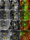
CiA-dependent non-cell autonomous accumulation of Diap-1 anterior to the MF. (a, a′ and a′′) cos2 and (b, b′ and b′′) cul-1 mosaic eye discs were labeled with anti-Diap-1 antibody. Mutant clones are marked by the absence of GFP. Clones located in or anterior to the MF (arrowhead) promote non-cell autonomous increase of Diap-1 levels (arrows). In contrast to cos2, mutant clones of cul-1 posterior to the MF contain reduced levels of Diap-1, indicating a requirement of cul-1 for Diap-1 regulation in addition to its role in Hh signaling. eF=ey-FLP. (c and d) Ectopic expression of CiA induces non-cell autonomous increase of Diap-1 levels in (c, c′ and c′′) eye and (d, d′ and d′′) wing discs (arrows). CiA-expressing clones are marked by the absence of GFP. hF=hs-FLP. Panels (ad) show the Diap-1 labelings only; panels (a′d′) show the GFP channel only; panels (a′′d′′) show the merge of Diap-1 and GFP. Genotypes: (a and b) y w ey-FLP; FRT42D cos2H29(a) or cul-1EX (b)/FRT42D P[ubi-GFP]. (c and d) y w hs-FLP; P[tubP>GFP>GAL4]; UAS-CiA. (> denotes FRT)
Figure 4
Figure 4
Non-cell autonomous upregulation of diap1 transcription by deregulated Hh signaling. (a and b) An enhancer trap insertion in the diap1 locus, diap1-lacZ (thj5c8), was used as a reporter for diap1 transcription. In both cos2 (a) and ptc (b) mosaics, ß-GAL levels are increased (yellow arrows in a′, a′′, b′ and b′′) anterior to the MF (white arrowheads) outside of the mutant clones (yellow outline in a′′ and b′′). However, there is also non-cell autonomous induction of diap1-lacZ immediately posterior to the MF (white arrow in a′). (c, c′, c′′) A cos2 mosaic eye disc labeled with anti-Ptc antibody. Genotypes: (a) ey-FLP; FRT42 cos2H29/FRT42 P[ubi-GFP]; thj5c8/+. (b) ey-FLP; FRT42 ptcX115/FRT42 P[ubi-GFP]; thj5c8/+. (c) ey-FLP; FRT42 cos2H29/FRT42 P[ubi-GFP]
Figure 5
Figure 5
Notch is required for cos2 suppression of GMR-hid and promotes Diap-1 accumulation non-cell autonomously. (ad) Heterozygosity of N (c) or Dl Ser (d) reverts the suppression of GMR-hid in cos2 mosaics (b), suggesting that Notch signaling is required for suppression of GMR-hid in cos2 mosaics. (e and e′) N protein strongly accumulates in cos2 clones (arrows) in or anterior to the MF (arrowhead). The N antibody (clone No. C17-9C6) was raised against the intracellular domain of N (NICD). (f and f′) cos2 induces N activity autonomously (red star) and non-cell autonomously (yellow arrows) anterior to the MF (arrowhead) as shown by an E(spl)m8-2.61-lacZ reporter expressing ß-GAL. (g, g′ and g′′) Ectopic expression of the active form of N, Nintra, induces Diap-1 levels non-cell autonomously immediately adjacent to Nintra-expressing clones (arrows), located in or anterior to the MF (arrowhead). Nintra-expressing clones are marked by the absence of the CD2 marker. (h) Statistical analysis of the suppression of GMR-hid by pka-C1 mosaics and pka-C1 Su(H) double mosaics. Eyes from GMR-hid flies are normalized to 100%. The suppression is partially reversed in double mosaics. For each bar, 10 eyes were averaged. **P-value ≤0.01. Genotypes: (a) y w GMR-hid ey-FLP/+; FRT42D P[w+]/CyO. (b) y w GMR-hid ey-FLP/+; FRT42D cos2H29/FRT42D P[w+]. (c) y w GMR-hid ey-FLP/N8; FRT42D cos2H29/FRT42D P[w+]. (d) y w GMR-hid ey-FLP/+; FRT42D cos2H29/FRT42D P[w+]; DlRevF10 SerRX82/+. (e and e′) y w ey-FLP; FRT42D cos2H29/FRT42D P[ubi-GFP]. (f and f′) y w ey-FLP; FRT42D cos2H29P[E(spl)m8-2.61-lacZ]/FRT42D P[ubi-GFP] P[E(spl)m8-2.61-lacZ]. (g, g′ and g′′) y w hs-FLP/+; P[Act5c>CD2>GAL4]; UAS-Nintra/+. (h) GMR-hid ey-FLP/+; P[ubi-GFP] FRT40/FRT40; GMR-hid ey-FLP/+; P[ubi-GFP] FRT40/pka-C1B3 FRT40; GMR-hid ey-FLP/+; P[ubi-GFP] FRT40/pka-C1B3 Su(H)Δ47 FRT40
Figure 6
Figure 6
Model of non-cell autonomous induction of Diap-1 by cells with ligand-independent deregulated Hh activity. Drawn are three cells at a clonal boundary in the MF, one mutant cell (cell A) with deregulated Hh activity (blue) and two non-mutant cells (cells B and C; white to light red). The mutant cell accumulates CiA, which promotes autonomous N activity (purple arrow) and stimulates Dl expression (green arrow). Autonomous N signaling releases an unknown extracellular factor that promotes transcription of diap1 in the first signal-receiving non-mutant cell, cell B. In the same non-mutant cell, Dl induces non-cell autonomous N activity, which in turn promotes transcription of diap1 in the second non-mutant cell, cell C. In this manner, cells with deregulated Hh signaling transmit increased apoptosis resistance to neighboring cells by upregulation of Diap-1. When the MF moves into clones of deregulated Hh activity, GMR-driven hid expression (red) will be inhibited by increased Diap-1 levels, resulting in suppression of the GMR-hid eye phenotype. The white-to-red color gradient in cells B and C indicates the approaching hid-expressing wave driven by GMR

Similar articles

Cited by

References

    1. Xu D, Woodfield SE, Lee TV, Fan Y, Antonio C, Bergmann A. Genetic control of programmed cell death (apoptosis) in Drosophila. Fly (Austin Tex) 2009;3:78–90. - PMC - PubMed
    1. Fuchs Y, Steller H. Programmed cell death in animal development and disease. Cell. 2011;147:742–758. - PMC - PubMed
    1. Kumar S. Caspase function in programmed cell death. Cell Death Differ. 2007;14:32–43. - PubMed
    1. Bergmann A. The role of ubiquitylation for the control of cell death in Drosophila. Cell Death Differ. 2010;17:61–67. - PMC - PubMed
    1. Bergmann A, Steller H. Apoptosis, stem cells, and tissue regeneration. Sci Signal. 2010;3:re8. - PMC - PubMed

Publication types