

A prospective study of preoperative computed tomographic angiographic mapping of free fibula osteocutaneous flaps for head and neck reconstruction
- PMID: 23018715
- PMCID: PMC3749731
- DOI: 10.1097/PRS.0b013e318262f115
A prospective study of preoperative computed tomographic angiographic mapping of free fibula osteocutaneous flaps for head and neck reconstruction
Abstract
Background: In designing an osteocutaneous fibula flap, poor planning, aberrant anatomy, or inadequate perforators may necessitate modification of the flap design, exploration of the contralateral leg, or additional flap harvest. The authors studied the predictive power of computed tomographic angiography in osteocutaneous fibula flap planning and execution.
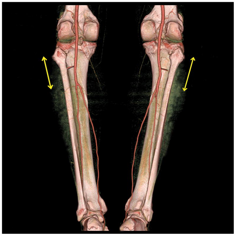
Methods: The authors studied a prospective cohort of 40 consecutive patients who underwent preoperative computed tomographic angiography mapping of the peroneal artery and its perforators and subsequent free fibula flap reconstruction of mandibular or maxillary defects. The authors compared their analysis of perforator anatomy, peroneal artery origin, and fibula length with intraoperative clinical findings.
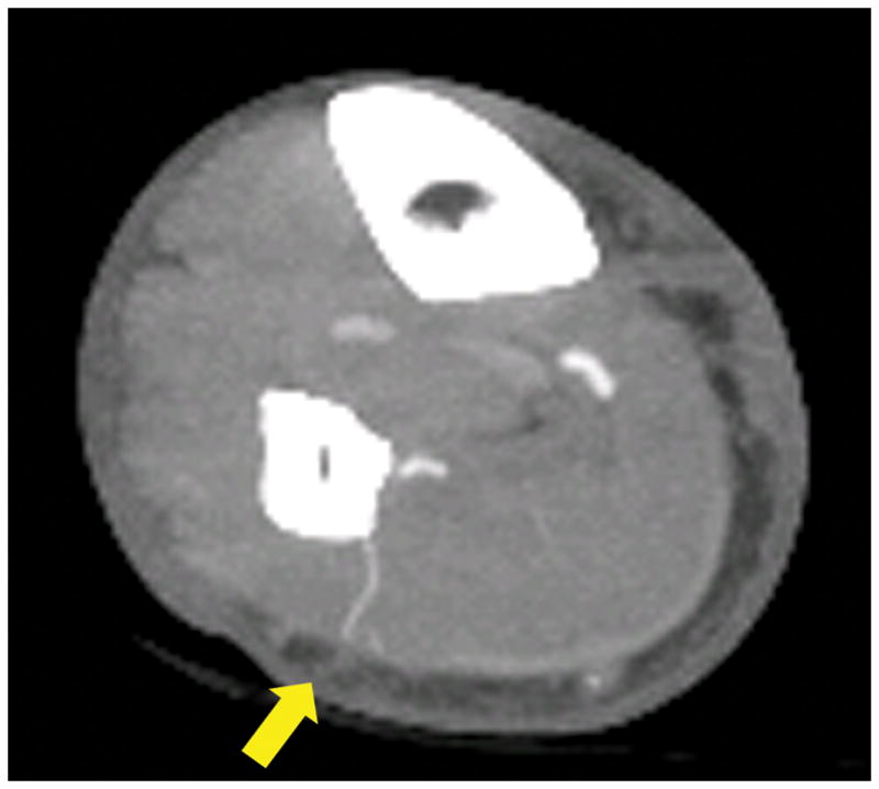
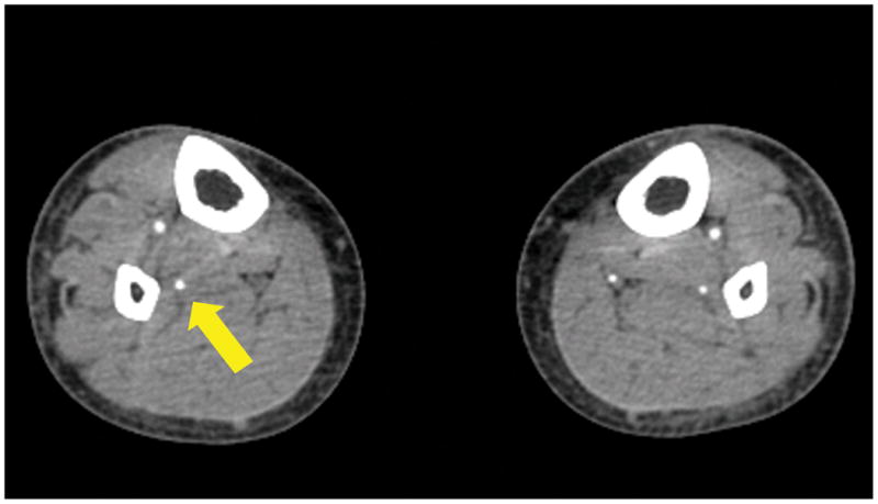
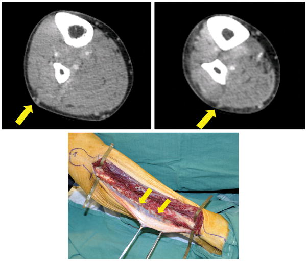
Results: Overall, computed tomographic angiography identified 94.9 percent of the cutaneous perforators found intraoperatively. Clinically, perforators were located an average of 8.7 mm from their predicted locations. The peroneal artery origin from the tibioperoneal trunk averaged 6.0 mm from its predicted location. The average length of the fibula differed from the predicted length by 8.0 mm. Computed tomographic angiography accurately predicted perforators as either septocutaneous or musculocutaneous 93.0 percent of the time. Perforator size was accurately predicted 66.7 percent of the time. Skin islands and osteotomies were modified in 25.0 percent of the cases on the basis of computed tomographic angiography findings. Two patients had hypoplastic posterior tibial arteries, prompting selection of the contralateral leg. There were no total flap or skin paddle losses.
Conclusions: Computed tomographic angiography accurately predicted the course and location of the peroneal artery and perforators; perforator size was less accurately estimated. Computed tomographic angiography provides valuable information to facilitate osteocutaneous fibula flap harvest.
Figures


References
-
- Wei FC, Chen HC, Chuang CC, Noordhoff MS. Fibular osteoseptocutaneous flap: anatomic study and clinical application. Plast Reconstr Surg. 1986;78:191–200. - PubMed
-
- Hidalgo DA. Fibula free flap: a new method of mandible reconstruction. Plast Reconstr Surg. 1989;84:71–80. - PubMed
-
- Cordeiro PG, Disa JJ, Hidalgo DA, Hu QY. Reconstruction of the mandible with osseous free flaps: a 10-year experience with 150 consecutive patients. Plast Reconstr Surg. 1999;104:1314–1320. - PubMed
-
- Taylor GI, Miller GD, Ham FJ. The free vascularized bone graft: a clinical extension of microvascular techniques. Plast Reconstr Surg. 1975;55:533–545. - PubMed
-
- Chen ZW, Yan W. The study and clinical application of the osteocutaneous flap of fibula. Microsurgery. 1983;4:11–16. - PubMed
