Review
. 2012 Dec;60(12):885-97.
doi: 10.1369/0022155412464638. Epub 2012 Sep 26.

An introduction to proteoglycans and their localization

Affiliations
Review

An introduction to proteoglycans and their localization

John R Couchman et al. J Histochem Cytochem. 2012 Dec.

Abstract

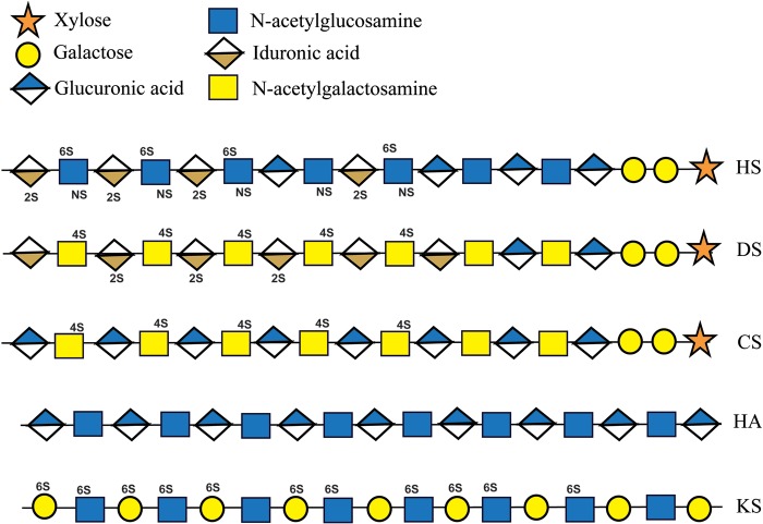
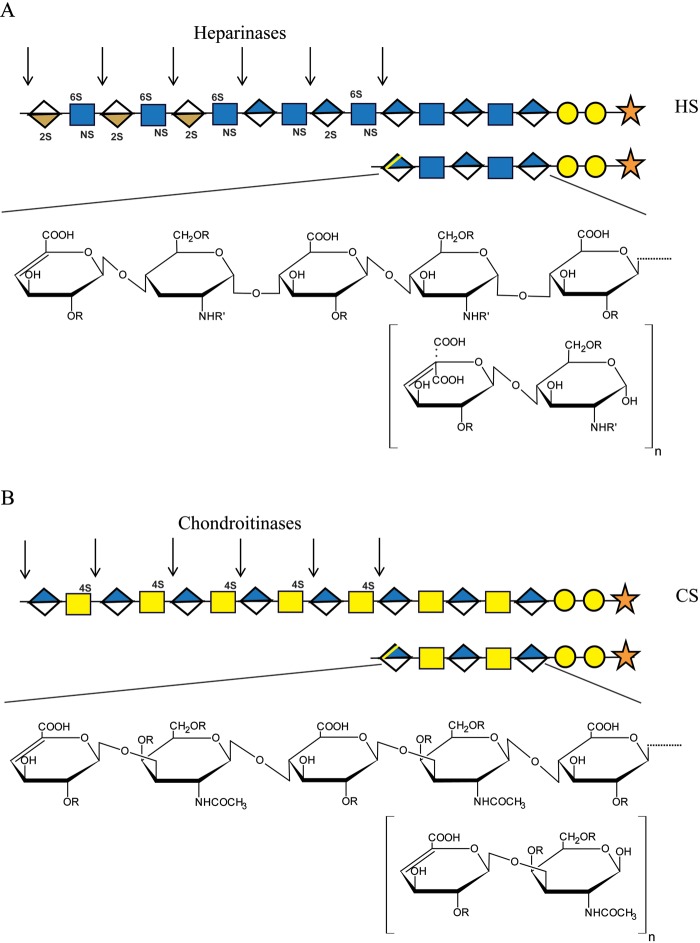
Proteoglycans comprise a core protein to which one or more glycosaminoglycan chains are covalently attached. Although a small number of proteins have the capacity to be glycanated and become proteoglycans, it is now realized that these macromolecules have a range of functions, dependent on type and in vivo location, and have important roles in invertebrate and vertebrate development, maintenance, and tissue repair. Many biologically potent small proteins can bind glycosaminoglycan chains as a key part of their function in the extracellular matrix, at the cell surface, and also in some intracellular locations. Therefore, the participation of proteoglycans in disease is receiving increased attention. In this short review, proteoglycan structure, function, and localizations are summarized, with reference to accompanying reviews in this issue as well as other recent literature. Included are some remarks on proteoglycan and glycosaminoglycan localization techniques, with reference to the special physicochemical properties of these complex molecules.


Conflict of interest statement

Declaration of Conflicting Interests: The authors declared no potential conflicts of interest with respect to the research, authorship, and/or publication of this article.

Figures

Figure 1.
Figure 1.
Composition of the glycosaminoglycans. Heparan sulfate (HS), chondroitin sulfate (CS), and dermatan sulfate (DS) all have a common stem oligosaccharide consisting of xylose and two galactose residues followed by a single glucuronic acid. Heparan sulfate (and the related highly sulfated heparin) then consists of a repeating disaccharide of N-acetylglucosamine and glucuronic acid. Subsequently, some of the N-acetylglucosamine is deacetylated and N-sulfated (NS). This usually occurs in blocks along the chain. These sulfated domains then undergo uronic acid epimerization, converting glucuronic acid to iduronic acid, some of which is then 2-O-sulfated (2S). Further modifications are 6-O and (rarely) 3-O sulfation of the hexosamine residues. In heparan sulfate, these modifications do not go to completion but culminate in domains of high sulfation (often referred to as NS domains) interspersed in regions of low or no sulfation (NA domains). Zones of intermediate sulfation can occur at the boundaries between these two extremes (NS/NA). Chondroitin and dermatan sulfates are similar: both consist of a repeating disaccharide of N-acetylgalactosamine and glucuronic acid. However, dermatan sulfate, by definition, has some of the uronic acid epimerized to iduronic acid by epimerase enzymes. Some of this iduronic acid can be sulfated at the 2-O position (2S). In the example shown here, both the CS and DS chains are sulfated at the 4-O position on the N-acetylgalactosamine (4S). However, chondroitin can be unsulfated or 6-O sulfated, and commonly a chain can contain more than one type of sulfation. Oversulfated forms of CS are known; for example, chondroitin sulfate E contains N-acetylgalactosamine sulfated at both the 4- and 6-O positions. Oversulfated forms of DS are also known. Hyaluronan (HA) comprises N-acetylglucosamine and glucuronic acid in a repeating disaccharide and may form chains of Mr = 1 × 10⁶ or more. It is synthesized at the cell surface and is therefore not modified by, for example, sulfation. Keratan sulfate (KS) consists of repeating disaccharides of galactose and N-acetylglucosamine, both of which can be 6-O sulfated.
Figure 2.
Figure 2.
Action of heparinases and chondroitinases on their respective glycosaminoglycans. These bacterial enzymes are eliminases and so, on cleavage of the polysaccharides, create an unsaturated uronic acid residue. One of these units will be present at the end of the carbohydrate “stub” remaining on each core protein. In addition, disaccharides or larger oligosaccharides will be released, each bearing a similar terminal unsaturated uronic acid. (A) There are three bacterial heparinases, I, II, and III, each of which has particular substrate specificity in terms of the heparan sulfate (HS) structure it will cleave. Heparinase III, for example, will cleave at unsulfated regions, and since the region proximal to the core protein is usually of low sulfation, this enzyme will pare back the polysaccharide close to the core protein. As a result of selectivity in heparan sulfate substrates that the three enzymes will cleave, oligosaccharides composed of varying numbers of disaccharide units will be released. To maximize heparan sulfate disaccharide cleavage products, it is common to use all three heparinases synchronously. For simplicity, the infrequent 3-O-sulfate modification of glucosamine is not shown. (B) Chondroitinase ABC will cleave chondroitin 4-sulfate (CS-A), dermatan sulfate (CS-B), and chondroitin 6-sulfate (CS-C), whereas chondroitinase ACII, for example, will not cleave dermatan sulfate. Chondroitin 4-sulfate is shown as a substrate in the model with symbols, but chondroitin may be unsulfated, 6-O sulfated, or a combination of these forms. However, in each case, a product with terminal unsaturated uronic acid will result from enzyme cleavage. R—H or SO3; R′—COCH3 or SO3.

References

    1. Afratis N, Gialeli C, Nikitovic D, Tsegenidis T, Karousou E, Theocharis A, Pavão MS, Tzanakakis GN, Karamanos NK. 2012. Glycosaminoglycans: key players in cancer biology and treatment. FEBS J. 279:1177–1197 - PubMed
    1. Allegretta M, Filmus J. 2011. Therapeutic potential of targeting glypican-3 in hepatocellular carcinoma. Anticancer Agents Med Chem. 11:543–548 - PMC - PubMed
    1. Arikawa-Hirasawa E, Le AH, Nishino I, Ho NC, Francomano CA, Govindraj P, Hassell JR, Devaney JM, Spranger J, Stevenson RE, et al. 2002. Structural and functional mutations of the perlecan gene cause Schwartz-Jampel syndrome, with myotonic myopathy and chondrodysplasia. Am J Hum Genet. 70:1368–1375 - PMC - PubMed
    1. Arikawa-Hirasawa E, Wilcox WR, Le AH, Sliverman N, Govindraj P, Hassell JR, Yamada Y. 2001. Dyssegmental dysplasia, Silverman-Handmaker type, is caused by functional null mutations of the perlecan gene. Nat Genet 27:431–434 - PubMed
    1. Arvatz G, Shafat I, Levy-Adam F, Ilan N, Voldavsky I. 2011. The heparanase system and tumor metastasis: is heparanase the seed and soil? Cancer Metastasis Rev. 30:253–268 - PubMed

Publication types