Review
. 2012 Dec;60(12):987-96.
doi: 10.1369/0022155412464376. Epub 2012 Sep 26.

The different roles of aggrecan interaction domains

Affiliations
Review

The different roles of aggrecan interaction domains

Anders Aspberg. J Histochem Cytochem. 2012 Dec.

Abstract

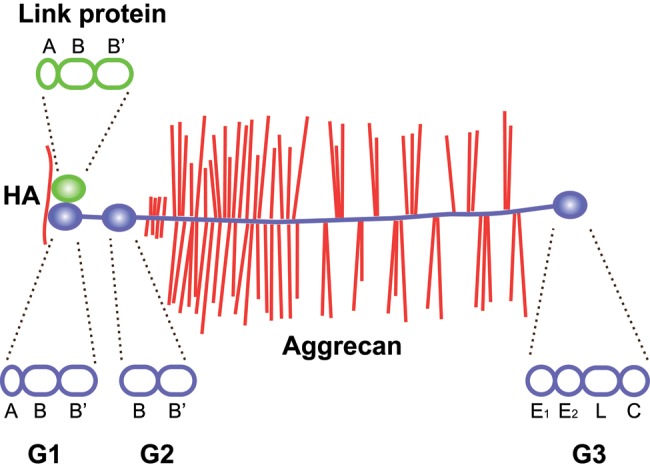
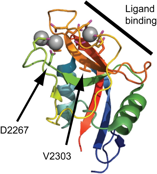
The aggregating proteoglycans of the lectican family are important components of extracellular matrices. Aggrecan is the most well studied of these and is central to cartilage biomechanical properties and skeletal development. Key to its biological function is the fixed charge of the many glycosaminoglycan chains, that provide the basis for the viscoelastic properties necessary for load distribution over the articular surface. This review is focused on the globular domains of aggrecan and their role in anchoring the proteoglycans to other extracellular matrix components. The N-terminal G1 domain is vital in that it binds the proteoglycan to hyaluronan in ternary complex with link protein, retaining the proteoglycan in the tissue. The importance of the C-terminal G3 domain interactions has recently been emphasized by two different human hereditary disorders: autosomal recessive aggrecan-type spondyloepimetaphyseal dysplasia and autosomal dominant familial osteochondritis dissecans. In these two conditions, different missense mutations in the aggrecan C-type lectin repeat have been described. The resulting amino acid replacements affect the ligand interactions of the G3 domain, albeit with widely different phenotypic outcomes.


Conflict of interest statement

Declaration of Conflicting Interests: The authors declared no potential conflicts of interest with respect to the research, authorship, and/or publication of this article.

Figures

Figure 1.
Figure 1.
Domain structure of aggrecan and link protein. Aggrecan binds hyaluronan through its N-terminal G1 domain in ternary complex with link proteins. The G1 domain and link proteins are homologous, with an immunoglobulin-like repeat (A) followed by two proteoglycan tandem repeats (B and B′). In aggrecan, but not in other lecticans, the G1 is followed by a second globular domain (G2), separated from the G1 by an extended interglobular domain. The G2 domain consists of two proteoglycan tandem repeats (B and B′). The central, and largest, part of aggrecan is the glycosaminoglycan attachment region, an extended protein stretch carrying keratan sulfate and chondroitin sulfate chains. This is followed by the C-terminal G3 domain, which consists of an epidermal growth factor (EGF) repeat (E1), a calcium-binding EGF repeat (E2), a C-type lectin domain (L), and a complement regulatory protein repeat (C). Hyaluronan (HA) is shown in red, aggrecan in blue (core protein) with red glycosaminoglycan chains, link protein in green. The structural repeat composition of each globular domain of link protein and aggrecan is shown above and below the two proteins, respectively, as indicated by dashed lines.
Figure 2.
Figure 2.
Schematic of lectican C-type lectin domain (CLD) interactions with tenascin and fibulin ligand proteins. The different interactions between lectican CLDs, tenascins, and fibulins are shown as lines. Line thicknesses indicate relative binding strengths, and affinities (KD) are indicated. The lectican proteoglycans (blue core proteins with red glycosaminoglycan chains) are shown attached to hyaluronan (HA, red line at the bottom of the figure) through their respective G1 domains in complex with Link protein (green). The G3 domain CLD ligands are shown in black at the top of the figure.
Figure 3.
Figure 3.
Lectican C-type lectin domain (CLD) binding sites on tenascins. Lectican CLD binding sites on tenascin-R and tenascin-C were mapped to fibronectin type III repeats 3 to 5 using panels of overlapping recombinant fragments. Affinities, determined by BIAcore surface plasmon resonance experiments, are in the low nanomolar range (KD:values for the aggrecan CLD are shown). The use of bacterially expressed tenascin proteins showed that the interactions were carbohydrate independent. The domain organization of the tenascins is shown with triangles for N-termini, spiral-filled circles for multimerization domains, diamonds for epidermal growth factor (EGF)–like repeats, ovals for fibronectin type III repeats, and hexagons for fibrinogen globules. Alternatively spliced fibronectin type III repeats are shadowed and shown with their insertion sites marked by dashed lines.
Figure 4.
Figure 4.
G3 domain-mediated organization of the aggrecan extracellular matrix. The model depicts how tenascin interaction through the lectican G3 domains may crosslink the proteoglycan aggregates and organize the extracellular matrix. Hyaluronan is shown in red (single red line forming the base), aggrecan in blue (core protein) with red glycosaminoglycan chains along the length of the core protein, link protein in green, and tenascin-C in black.
Figure 5.
Figure 5.
Disease-linked missense mutations in the aggrecan C-type lectin domain (CLD). The aggrecan C-type lectin domain structure determined by X-ray crystallography (Protein Data Bank ID: 1TDQ) is shown as a cartoon model. The coordinated calcium ions are shown as gray spheres. The side chains of amino acid residues coordinating the calcium ions or mutated in human disease are shown as sticks. The CLD binding surface for tenascin-R is marked by a thick line in the upper right corner of the cartoon. The amino acid residues mutated in spondyloepimetaphyseal dysplasia aggrecan type (D2267) and familial osteochondritis dissecans (V2303) are indicated by arrows.

References

    1. Aspberg A, Adam S, Kostka G, Timpl R, Heinegård D. 1999. Fibulin-1 is a ligand for the C-type lectin domains of aggrecan and versican. J Biol Chem. 274:20444–20449 - PubMed
    1. Aspberg A, Binkert C, Ruoslahti E. 1995. The versican C-type lectin domain recognizes the adhesion protein tenascin-R. Proc Natl Acad Sci U S A. 92:10590–10594 - PMC - PubMed
    1. Aspberg A, Miura R, Bourdoulous S, Shimonaka M, Heinegård D, Schachner M, Ruoslahti E, Yamaguchi Y. 1997. The C-type lectin domains of lecticans, a family of aggregating chondroitin sulfate proteoglycans, bind tenascin-R by protein-protein interactions independent of carbohydrate moiety. Proc Natl Acad Sci U S A. 94:10116–10121 - PMC - PubMed
    1. Bonaventure J, Kadhom N, Cohen-Solal L, Ng KH, Bourguignon J, Lasselin C, Freisinger P. 1994. Reexpression of cartilage-specific genes by dedifferentiated human articular chondrocytes cultured in alginate beads. Exp Cell Res. 212:97–104 - PubMed
    1. Cavanagh JA, Tammen I, Windsor PA, Bateman JF, Savarirayan R, Nicholas FW, Raadsma HW. 2007. Bulldog dwarfism in Dexter cattle is caused by mutations in ACAN. Mamm Genome. 18:808–814 - PubMed

Publication types