Review
. 2013 Feb;51(2):75-82.
doi: 10.1002/dvg.22349. Epub 2012 Oct 16.

Determining how defects in connexin43 cause skeletal disease

Affiliations
Review

Determining how defects in connexin43 cause skeletal disease

Quynh V Ton et al. Genesis. 2013 Feb.

Abstract

Gap junction channels mediate direct cell-cell communication via the exchange of second messengers, ions, and metabolites from one cell to another. Mutations in several human connexin (cx) genes, the subunits of gap junction channels, disturb the development and function of multiple tissues/organs. In particular, appropriate function of Cx43 is required for skeletal development in all vertebrate model organisms. Importantly, it remains largely unclear how disruption of gap junctional intercellular communication causes developmental defects. Two groups have taken distinct approaches toward defining the tangible molecular changes occurring downstream of Cx43-based gap junctional communication. Here, these strategies for determining how Cx43 modulates downstream events relevant to skeletal morphogenesis were reviewed.


Figures

Figure 1
Figure 1
Gap junction channels connect the cytoplasm of adjacent cells. Each connexin contains four transmembrane-spanning domains, with both the amino and carboxy ends located in the cytoplasm. Six connexins comprise a connexon, or hemichannel. Two connexons, one from each cell, dock together at the plasma membrane to make a single gap junction channel. IL, intracellular loop; EL, extracellular loop; TM, transmembrane-spanning domain.
Figure 2
Figure 2
The in vitro system used to evaluate Cx43-dependent regulation of gene expression. The reporter construct contains the osteocalcin (OCN) promoter followed by the coding sequence for the luciferase gene. The level of luciferase transcription is determined by quantitating the amount of luciferase activity. Osteoblast cell lines transfected with this reporter demonstrate that luciferase activity is dependent on the level of Cx43-based function.
Figure 3
Figure 3
Model showing how gap junctions may amplify signals originating from growth-factor mediated cascades. A primary signal originates from a growth factor-receptor interaction that can initiate a response/change in gene expression in cell one (not drawn). At the same time, second messengers resulting from the primary signal transverse gap junction channels to initiate a similar change in gene expression in cell two. This is the secondary signal. Described Cx43-dependent changes include Ras/ERK (triangle) mediated phosphorylation of Sp1 (Stains and Civitelli, 2005) and PKCδ (diamond) mediated phosphorylation of Runx2 (Lima et al., 2009). PKCδ may also independently activate ERK (Niger et al., 2012). Phosphorylated Sp1 and Runx2 favor transcription of certain osteoblast genes.
Figure 4
Figure 4
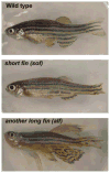
Fin length mutants exhibit defects in skeletal morphogenesis. Top: wild-type zebrafish. Middle: sof b123 mutant. Bottom: alf dty86 mutant.
Figure 5
Figure 5
Cx43 influences cell proliferation and joint formation by regulating the Sema3d pathway. (a) Expression of the sema3d gene is regulated by cx43. The Sema3d protein is secreted and interacts with its putative receptors, Nrp2a and PlxnA3, to regulate cell proliferation and joint formation respectively. (b) Compartmental expression of the genes in the Cx43 pathway. The cx43 mRNA is up-regulated in the proliferating cells of the blastema (red), while sema3d is up-regulated in the lateral skeletal precursor cells and basal layer of the epidermis (green). The nrp2a gene is expressed in the distal blastema (yellow), where it may regulate cell proliferation in the blastemal cells. The plxna3 gene is expressed in the skeletal precursor cells (blue), where it may regulate joint formation. This figure was originally published in Ton and Iovine, 2012, and is reprinted here with permission.
Figure 6
Figure 6
Examples of how Cx43-dependent GJIC may influence gene expression in the regenerating fin. (a) Up-regulation of cx43 in the blastema (red) may lead to the secretion of a growth factor that can interact with its receptor located on adjacent skeletal precursor cells (green), leading to increased sema3d expression. (b) Heterotypic gap junctions may exist between cells of the blastema and skeletal precursor cells, permitting the direct exchange of secondary messengers. These second messengers may influence sema3d expression.

References

    1. Akimenko MA, Mari-Beffa M, Becerra J, Geraudie J. Old questions, new tools, and some answers to the mystery of fin regeneration. Dev Dyn. 2003;226:190–201. - PubMed
    1. Bivi N, Lezcano V, Romanello M, Bellido T, Plotkin LI. Connexin43 interacts with βarrestin: a pre-requisite for osteoblast survival induced by parathyroid hormone. J Cell Biochem. 2011;112:2920–2930. - PMC - PubMed
    1. Bivi N, Condon KW, Allen MR, Farlow N, Passeri G, Brun LR, Rhee Y, Bellido T, Plotkin LI. Cell autonomous requirement of connexin 43 for osteocyte survival: consequences for endocortical resorption and periosteal bone formation. J Bone Miner Res. 2012;27:374–89. - PMC - PubMed
    1. Brown AM, Fisher S, Iovine MK. Osteoblast maturation occurs in overlapping proximal-distal compartments during fin regeneration in zebrafish. Dev Dyn. 2009;238:2922–2928. - PMC - PubMed
    1. Chung DJ, Castro CH, Watkins M, Stains JP, Chung MY, Szejnfeld VL, Willecke K, Theis M, Civitelli R. Low peak bone mass and attenuated anabolic response to parathyroid hormone in mice with an osteoblast-specific deletion of connexin43. J Cell Sci. 2006;119:4187–98. - PubMed

Publication types

LinkOut - more resources