. 2012 Dec 15;303(12):F1629-40.
doi: 10.1152/ajprenal.00246.2012. Epub 2012 Sep 26.

Null mutations at the p66 and bradykinin 2 receptor loci induce divergent phenotypes in the diabetic kidney

Affiliations

Null mutations at the p66 and bradykinin 2 receptor loci induce divergent phenotypes in the diabetic kidney

Himanshu Vashistha et al. Am J Physiol Renal Physiol. .

Abstract

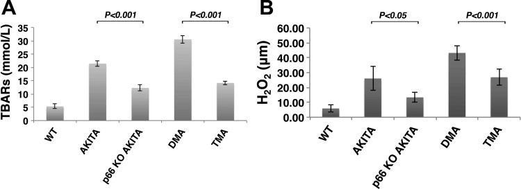
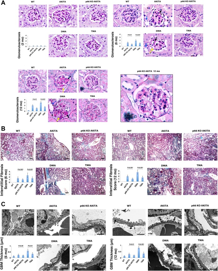
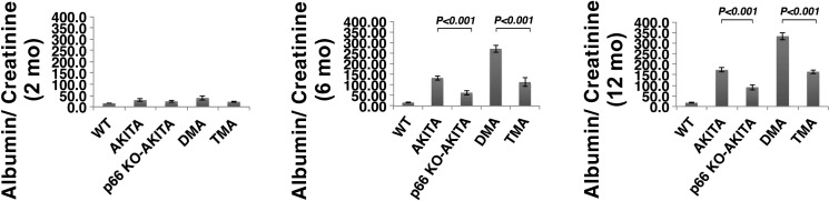
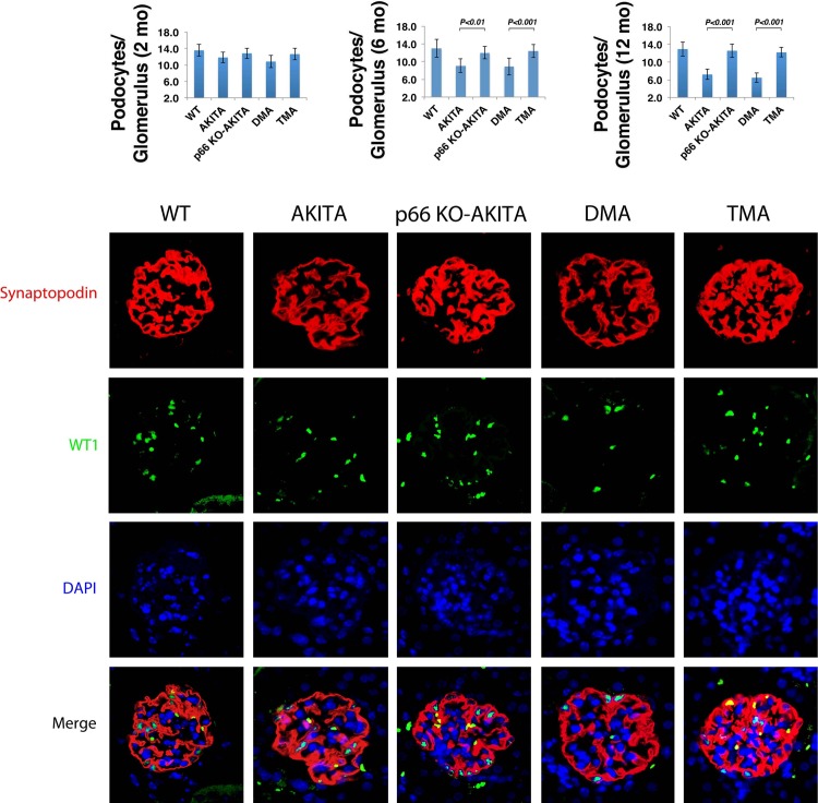
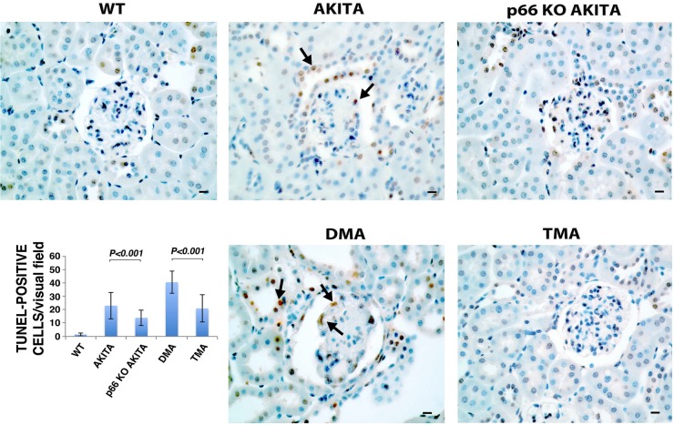
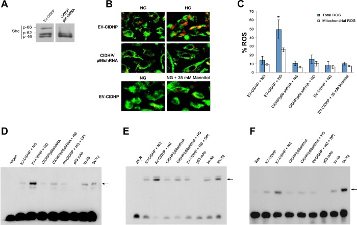
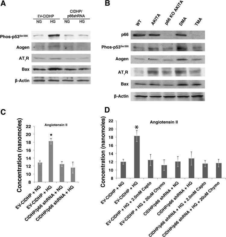
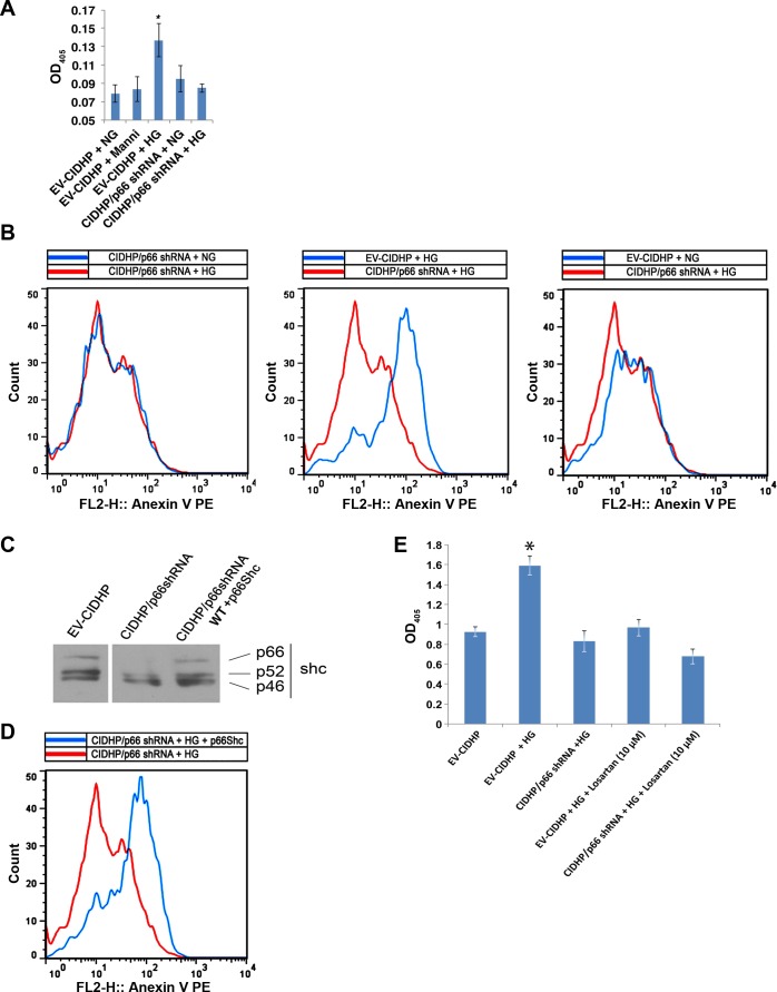
Candidate genes have been identified that confer increased risk for diabetic glomerulosclerosis (DG). Mice heterozygous for the Akita (Ins2(+/C96Y)) diabetogenic mutation with a second mutation introduced at the bradykinin 2 receptor (B2R(-/-)) locus express a disease phenotype that approximates human DG. Src homology 2 domain transforming protein 1 (p66) controls mitochondrial metabolism and cellular responses to oxidative stress, aging, and apoptosis. We generated p66-null Akita mice to test whether inactivating mutations at the p66 locus will rescue kidneys of Akita mice from disease-causing mutations at the Ins2 and B2R loci. Here we show null mutations at the p66 and B2R loci interact with the Akita (Ins2(+/C96Y)) mutation, independently and in combination, inducing divergent phenotypes in the kidney. The B2R(-/-) mutation induces detrimental phenotypes, as judged by increased systemic and renal levels of oxidative stress, histology, and urine albumin excretion, whereas the p66-null mutation confers a powerful protection phenotype. To elucidate the mechanism(s) of the protection phenotype, we turned to our in vitro system. Experiments with cultured podocytes revealed previously unrecognized cross talk between p66 and the redox-sensitive transcription factor p53 that controls hyperglycemia-induced ROS metabolism, transcription of p53 target genes (angiotensinogen, angiotensin II type-1 receptor, and bax), angiotensin II generation, and apoptosis. RNA-interference targeting p66 inhibits all of the above. Finally, protein levels of p53 target genes were upregulated in kidneys of Akita mice but unchanged in p66-null Akita mice. Taken together, p66 is a potential molecular target for therapeutic intervention in DG.


Figures

Fig. 1.
Fig. 1.
The p66 is necessary for hyperglycemia (HG)-induced reactive oxygen species (ROS) metabolism. A: plasma levels of thiobarbituric acid reactive substances (TBARs) in Akita diabetic mice. WT, wild type; KO, knockout. Data are presented as means ± SD; n = 5 in each group. P < 0.001, Akita vs. p66KO-Akita; P < 0.001, double mutant Akita (DMA) vs. triple mutant Akita (TMA). B: H2O2 content in kidney tissue from Akita diabetic mice. Data are presented as means ± SD; n = 5 in each group; P < 0.05, Akita vs. p66KO-Akita; P < 0.001, DMA vs. TMA.
Fig. 2.
Fig. 2.
p66 is necessary for expression of disease phenotype(s) in kidneys of Akita mice. A: periodic acid-Schiff (PAS) staining of kidney sections. Ins2 mutation alone increases accumulation of PAS-positive matrix at 6 mo (right) and 12 mo of age (bottom), which is enhanced at 6 and 12 mo by the absence of the B2R. The p66-null mutation attenuates renal histological changes at corresponding intervals. Nodular PAS-positive matrix (black arrow), mesangiolysis (yellow arrow) tubular dilation (blue arrow) and microaneurysm (arrowhead). Scale bar = 10 μm. Histologic analysis of glomerulosclerosis by semiquantitative morphometric analysis (inset). Results are presented as means ± SD; n = 5 in each group. P < 0.001, Akita vs. p66KO-Akita; P < 0.001, DMA vs. TMA. B: trichrome staining of renal cortex. DMA mice show increase interstitial fibrosis at 6 (left) and 12 mo of age (right), whereas p66-null mutation attenuates interstitial fibrosis at corresponding intervals. Scale bar = 50 μm. Histologic analysis of interstitial fibrosis assessed by semi-quantitative evaluation (inset). Results are presented as means ± SD; n = 5 in each group. P < 0.001, Akita vs. p66 KO Akita (12 mo); P < 0.001, DMA vs. TMA. C: electron micrographs of glomeruli. Akita and DMA mice at 6 (left) and 12 (right) mo of age show thickening of glomerular basement membrane (GBM) and foot process effacement. p66KO-Akita and TMA show minimum GBM thickening and intact foot processes at corresponding interval. Scale bar = 1 μm. Inset: quantitative analysis of GBM thickness. Results are presented as means ± SD; n = 5 in each group. P < 0.001, p66KO-Akita vs. Akita; P < 0.001, DMA vs. TMA.
Fig. 3.
Fig. 3.
The p66 null mutation attenuates urine albumin excretion (UAE). Akita mice at 2 mo (left), 6 mo (middle), and 12 mo of age (right). Results are presented as means ± SD; n = 5 in each group. P < 0.001, Akita vs. p66 KO Akita; P < 0.001, DMA vs. TMA.
Fig. 4.
Fig. 4.
Podocyte number/glomerulus. WT-1 expressing nuclei were counted in kidneys of Akita mice at 2 mo (left), 6 mo (middle), and 12 mo of age (right). Results are presented as means ± SD; n = 5 in each group; 30 glomeruli from each mouse were analyzed. P 0.01, p66 KO Akita vs. Akita (6 mo); P 0.001, p66 KO Akita vs. Akita (12 mo), DMA vs. TMA (6 and 12 mo). Confocal images of glomeruli showing podocyte specific marker synaptopodin (red); podocyte nuclear marker WT-1 (green); nuclear counterstain DAPI (blue) in kidneys sections at 12 mo of age.
Fig. 5.
Fig. 5.
Detection of apoptosis by terminal transferase-mediated deoxyuridine triphosphate nick end labeling (TUNEL) assay. Representative images of TUNEL staining in kidney sections of WT and Akita mice. Arrows indicate TUNEL-positive cells. Scale bar = 10 μm. Quantitative analysis of TUNEL-positive nuclei. Results shown are presented as means ± SD; n = 5. P < 0.01, Akita vs. p66KO-Akita; P < 0.01, DMA vs. TMA.
Fig. 6.
Fig. 6.
The p66 redox signals activate p53 DNA binding. A: immunoblot analysis of ShcA isoforms in conditionally immortalized differentiated human podocytes (CIDHP) stably transfected with p66shRNA. B: CIDHP/p66shRNA shows inhibition of HG-induced ROS metabolism. C: quantitative of ROS in empty vector (EV)-CIDHP and CIDHP/p66 shRNA cells. *P < 0.01. EMSA showing p53 binding to the consensus sequence at the promoter of Aogen (D), angiotensin II type 1 receptor (AT1R; E), and bax (F). Nuclear extracts from SV-T2 cells overexpressing p53 were used as a positive control. Arrow indicates position of p53 specific band. Aogen indicates Aogen probe; AT1R indicates AT1R probe; Bax indicates bax probe; each case in the absence of nuclear extracts. Specificity was established by documenting incubation with monoclonal p53 antibody (p53 mAb) inhibits the p53 band complex whereas irrelevant antibody (Irr-p53) was without effect. EV-CIDHP at HG show increase in the intensity of p53 specific band complex for Aogen, AT1R, and bax. EV-CIDHP maintained at HG, in the presence of free radical scavenger DPI, show no increase in the intensity of the p53 band complex for Aogen, AT1R, and bax. Results shown are representative of 4 independent experiments.
Fig. 7.
Fig. 7.
The p66 is necessary for expression of p53 target genes. A: effect of p66-shRNA on expression of p53 target genes. Representative immunoblot analysis of Aogen, AT1R, and bax protein levels. Results shown are representative of 4 independent experiments. B: effect of p66-null mutation on renal expression of p53 and p53 target genes. Represenative immunoblot analysis of kidney from WT and Akita mice at 12 mo of age. Results shown are representative of 4 independent experiments. C: effect of p66-shRNA on synthesis and release of angiotensin II. D: effect of ACE and non-ACE inhibitors on HG induced angiotensin II generation. Data are presented as means ± SD and represent 4 independent experiments. *P ≤ 0.01.
Fig. 8.
Fig. 8.
The p66 and angiotensin II are necessary for HG-induced apoptosis. A: histone associated DNA fragments were quantified and presented as optical density at 405 nm (OD405). Results are presented as means ± SD and represent 4 independent experiments. *P < 0.01. B: Representative flow cytometric analysis showing detection of apoptosis by annexin V binding. Results shown are representative of 4 independent experiments. C: representative immunoblot analysis showing expression of WT p66ShcA in CIDHP/p66shRNA. Results shown are representative of 4 independent experiments. D: flow cytometric analysis of annexin V binding with reconstituted CIDHP/p66shRNA. Results shown are representative of 4 independent experiments. E: losartan inhibits HG-induced apoptosis. Results are presented as means ± SD and represent 3–4 independent experiments. *P < 0.01.

References

    1. Brenner BM, Cooper ME, de Zeeuw D, Keane WF, Mitch WE, Parving HH, Remuzzi G, Snapinn SM, Zhang Z, Shahinfar S. Effects of losartan on renal and cardiovascular outcomes in patients with type 2 diabetes and nephropathy. N Engl J Med 345: 861–869, 2001 - PubMed
    1. Brenner BM. Remission of renal disease: recounting the challenge, acquiring the goal. J Clin Invest 110: 1753–1758, 2002 - PMC - PubMed
    1. Camici GG, Schiavoni M, Francia P, Bachschmid M, Martin-Padura I, Hersberger M, Tanner FC, Pelicci P, Volpe M, Anversa P, Luscher TF, Cosentino F. Genetic deletion of p66(Shc) adaptor protein prevents hyperglycemia-induced endothelial dysfunction and oxidative stress. Proc Natl Acad Sci USA 104: 5217–5222, 2007 - PMC - PubMed
    1. Chintapalli J, Yang S, Opawumi D, Goyal SR, Shamsuddin N, Malhotra A, Reiss K, Meggs LG. Inhibition of wild-type p66ShcA in mesangial cells prevents glycooxidant-dependent FOXO3a regulation and promotes the survival phenotype. Am J Physiol Renal Physiol 292: F523–F530, 2007 - PubMed
    1. Durvasula RV, Shankland SJ. Activation of a local renin angiotensin system in podocytes by glucose. Am J Physiol Renal Physiol 294: F830–F839, 2008 - PubMed

Publication types

MeSH terms

Substances