. 2012 Nov 16;287(47):39789-99.
doi: 10.1074/jbc.M112.383000. Epub 2012 Sep 27.

Activation of autoreactive B cells by endogenous TLR7 and TLR3 RNA ligands

Affiliations

Activation of autoreactive B cells by endogenous TLR7 and TLR3 RNA ligands

Nathaniel M Green et al. J Biol Chem. .

Abstract

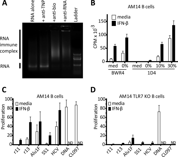
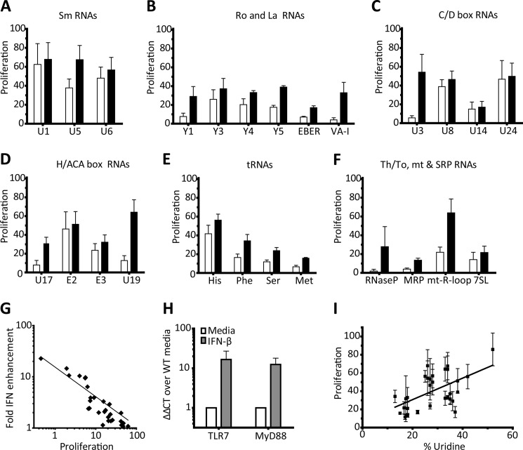
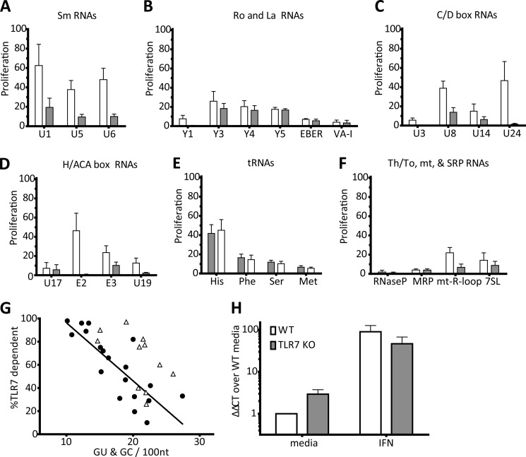
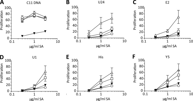
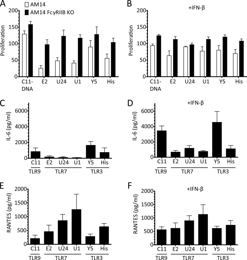
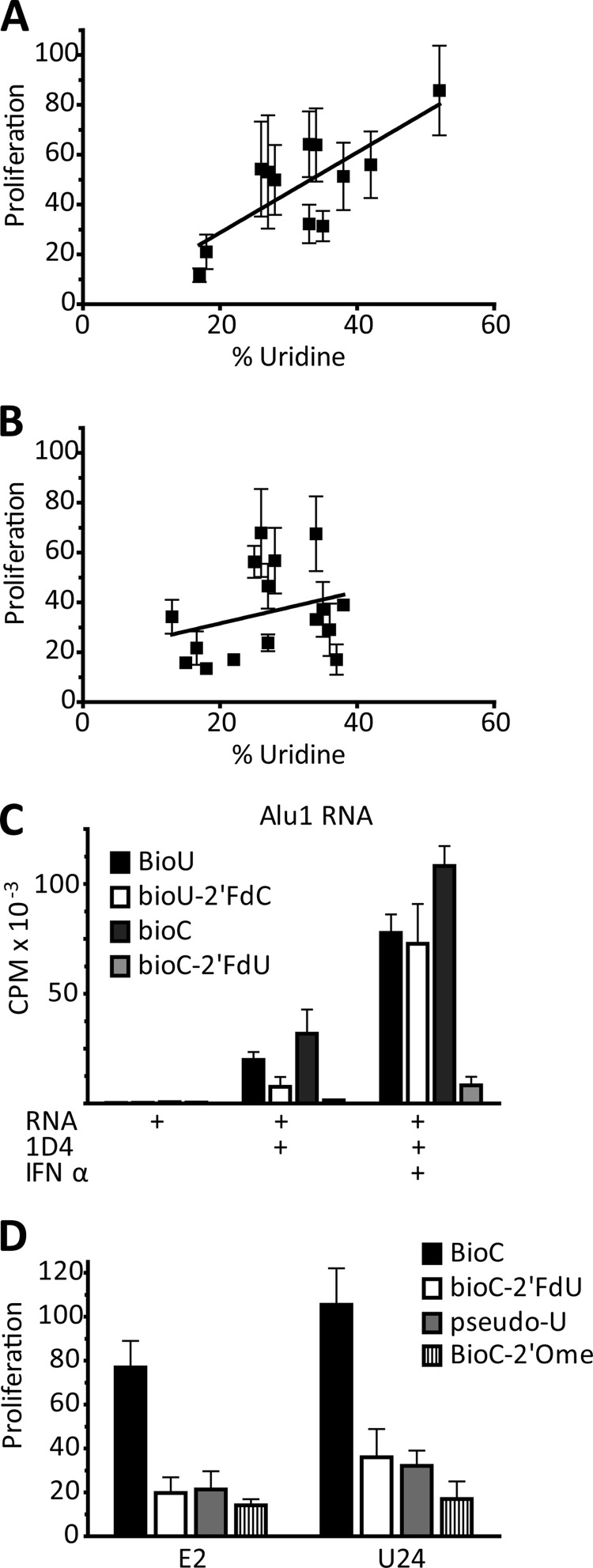
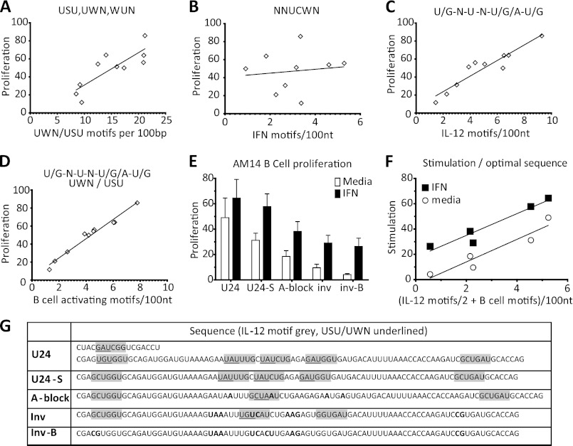
The key step in the activation of autoreactive B cells is the internalization of nucleic acid containing ligands and delivery of these ligands to the Toll-like Receptor (TLR) containing endolysosomal compartment. Ribonucleoproteins represent a large fraction of autoantigens in systemic autoimmune diseases. Here we demonstrate that many uridine-rich mammalian RNA sequences associated with common autoantigens effectively activate autoreactive B cells. Priming with type I IFN increased the magnitude of activation, and the range of which RNAs were stimulatory. A subset of RNAs that contain a high degree of self-complementarity also activated B cells through TLR3. For the RNA sequences that activated predominantly through TLR7, the activation is proportional to uridine-content, and more precisely defined by the frequency of specific uridine-containing motifs. These results identify parameters that define specific mammalian RNAs as ligands for TLRs.


Figures

FIGURE 1.
FIGURE 1.
Uridine-rich RNAs activate AM14 B cells, when delivered as ICs, through a TLR7-dependent manner. A, in vitro transcribed bio-Alu1 RNA, alone or premixed with the indicated mAbs, was electrophoresed in a 1% agarose gel and subsequently visualized with ethidium bromide. B, AM14 B cells were stimulated with BWR4 or 1D4 alone (med) or premixed with bio-Alu1 RNA transcribed in the presence of 0, 10, or 30% biotinylated cytodine bases, in the absence □ or presence ■ of IFN-β. Proliferation was quantified by [3H]thymidine incorporation. Average ± S.E. is shown, n = 3. C and D, AM14 B cells, or AM14 TLR7-deficient B cells were stimulated with 1D4 premixed with the indicated bio-RNAs (with 8 biotins for every 100 nt) or stimulated with 1D4 and bio-DNA, or the small molecule ligand CL097 and in the absence □ or presence ■ of IFN-β. Proliferation is measured by [3H]thymidine uptake, and plotted as the percent of the response to CpG-B. Average ± S.E. is shown, n = 3.
FIGURE 2.
FIGURE 2.
The response of AM14 B cells to endogenous, RNP-associated RNA sequences is variably enhanced by type I IFN. A–F, AM14 B cells were stimulated with 1D4 or premixed with the indicated bio-RNAs, in the absence □ or presence ■ of IFN-β. Proliferation was quantified by [3H]thymidine incorporation, and is shown as the percent of the response to CpG-B. Average ± S.E. is shown, A and C, n = 4, B and D–F n = 3. G, proliferation of the samples from A–F in the absence of interferon plotted against the fold enhancement in the presence of IFN-β. Log-log regression line Y = 10(−0.590×log X +1.164), R2 = 0.8543. H, qRT-PCR for TLR7 and MyD88 from resting and 1 h IFN-primed AM14 B cells. Data represented as ΔΔCT fold change over media, relative to GAPDH. Average ± S.E. is shown, n = 5. I, proliferation of the samples from A–F in the presence of IFN, plotted against % of uridine content in each fragment. Linear regression line, R2 = 0.3206, p = 0.0011.
FIGURE 3.
FIGURE 3.
Endogenous RNP-associated RNA sequences with regions of sequence complementarity can activate AM14 B cells independently of TLR7. A–F, AM14 □ or AM14 TLR7-deficient ■ B cells were stimulated with 1D4 premixed with the indicated bio-RNAs. Proliferation is plotted as the percent of the response to CpG-B. Average ± S.E. is shown, A and C, n = 4, B and D–F, n = 3. G, percent of proliferation dependent of TLR7 from A–F, plotted against the number of base-paired guanines predicted by MFOLD. Stimulatory RNAs (●) were compared for the linear regression, R2 = 0.6575, p < 0.0001, weakly stimulatory (<10% Proliferation on WT) RNAs (Δ) were not included in this analysis. H, qRT-PCR for TLR3 from resting and 1 h IFN-primed AM14 □ or AM14 TLR7 KO ■ B cells. Data represented as ΔΔCT fold change over media, relative to GAPDH. Average ± S.E. is shown, n = 3.
FIGURE 4.
FIGURE 4.
Endogenous RNP-associated RNA sequences with regions of sequence complementarity can activate B cells through TLR3. A–F, WTΔ, TLR7 KO □, TLR3/7 dKO ▾, or unc93b3d ο B cells were stimulated with the indicated Fab ICs. Proliferation was quantified by [3H]thymidine incorporation and plotted as the percent of the response to CpG-B. Average ± S.E. is shown, n = 7.
FIGURE 5.
FIGURE 5.
TLR3-dependent RNAs make IL-6 independently of IFN priming. A and B, proliferation of AM14 □ and AM14 FcγRIIB KO ■ B cells were stimulated with with 1D4 or premixed with the indicated bio-RNAs, in the absence (A) or presence (B) of IFN-β. Proliferation was quantified by [3H]thymidine incorporation, and is shown as the percent of the response to CpG-B. Average ± S.E. is shown, n = 4. C–F, cytokine production of AM14 FcγRIIB KO B cells in the absence (C and E) or presence (D and F) of IFN-β. The TLRs through which the given ligands stimulate are noted under the name of the nucleic acid. Cytokine production was measured by ELISA, and the average ± S.E. of four independent experiments is shown.
FIGURE 6.
FIGURE 6.
Uridines are an essential determining factor in RNA activation through TLR7. A, proliferation in the presence of IFN of the samples from Fig. 3, A–F, plotted against % uridine content of the fragment; analysis includes RNAs that are >75% TLR7 dependent. Linear regression, R2 = 0.5840, p = 0.0038. B, proliferation in the presence of IFN of the samples from Fig. 3, A–F, plotted against % uridine content of the fragment; analysis includes RNAs that are <75% TLR7 dependent for stimulation. Linear regression line, R2 = 0.02927, p = 0.5115. C, Bio-Alu 1 RNA ICs activation of AM14 B cells ± IFN-β; RNAs transcribed with BioU or Bio-C, and/or 2′FdC or 2′FdU as indicated. Proliferation is measured by [3H]thymidine incorporation. Average ± S.E. is shown, n = 3. D, Bio-E2 and Bio-U24 RNA IC activation of AM14 B cells; RNAs transcribed with Bio-C and either 2′-FdU, pseudouridine (pseudo-U), or 2′-O-methyl-uridine (2′Ome) substitutions. Proliferation is plotted as the percent of the response to CpG-B. Average ± S.E. is shown, n = 3.
FIGURE 7.
FIGURE 7.
A specific uridine-containing motif determines the stimulatory capacity of RNA ligands for TLR7. A, proliferation, in the presence of IFN, of the >90% TLR7-dependent samples from Fig. 3, A–F plotted against the frequency of USU/UWN 3-mers. Linear regression, R2 = 0.6771, p = 0.0035. B, proliferation, in the presence of IFN, of the >90% TLR7 dependent samples from Fig. 3, A–F plotted against NNUCWN (IFN) motifs. Linear regression, R2 = 0.01623, p = 0.7440. C, proliferation, in the presence of IFN, of the >90% TLR7-dependent samples from Fig. 3, A–F plotted against frequency of KNUNDK (IL-12) motif. Linear regression, R2 = 0.8838, p < 0.0001. D, proliferation, in the presence of IFN, of the >90% TLR7-dependent samples from Fig. 3, A–F plotted against frequency of KNUNDK & USU/UWN (B cell activating) motif. Linear regression, R2 = 0.9710, p < 0.0001. E, U24-bio RNA, and sequence modifications, with 1D4 stimulation of AM14 B cells, in the absence □ or presence ■ of IFN-β. Proliferation is measured by [3H]thymidine uptake, and plotted as the percent of the response to CpG-B. Average ± S.E. is shown, n = 3. F, proliferation from E plotted against the frequency of B cell stimulating motifs + half the IL-12 motifs that do not fulfill the USU/UWN motif. Linear regression for media: R2 = 0.8964, p = 0.0146. Linear regression for IFN: R2 = 0.9242, p = 0.0091. G, sequences of RNAs analyzed in E and F.

References

    1. Tan E. M., Cohen A. S., Fries J. F., Masi A. T., McShane D. J., Rothfield N. F., Schaller J. G., Talal N., Winchester R. J. (1982) The 1982 revised criteria for the classification of systemic lupus erythematosus. Arthritis Rheum. 25, 1271–1277 - PubMed
    1. Lerner M. R., Steitz J. A. (1979) Antibodies to small nuclear RNAs complexed with proteins are produced by patients with systemic lupus erythematosus. Proc. Natl. Acad. Sci. U.S.A. 76, 5495–5499 - PMC - PubMed
    1. Harris M. L., Rosen A. (2003) Autoimmunity in scleroderma: the origin, pathogenetic role, and clinical significance of autoantibodies. Current Opin. Rheumatol. 15, 778–784 - PubMed
    1. Levine S. M., Raben N., Xie D., Askin F. B., Tuder R., Mullins M., Rosen A., Casciola-Rosen L. A. (2007) Novel conformation of histidyl-transfer RNA synthetase in the lung: the target tissue in Jo-1 autoantibody-associated myositis. Arthritis Rheum. 56, 2729–2739 - PubMed
    1. Lau C. M., Broughton C., Tabor A. S., Akira S., Flavell R. A., Mamula M. J., Christensen S. R., Shlomchik M. J., Viglianti G. A., Rifkin I. R., Marshak-Rothstein A. (2005) RNA-associated autoantigens activate B cells by combined B cell antigen receptor/Toll-like receptor 7 engagement. J. Exp. Med. 202, 1171–1177 - PMC - PubMed

Publication types

LinkOut - more resources