. 2012 Sep 25;109(39):15769-74.
doi: 10.1073/pnas.1204357109. Epub 2012 Sep 10.

Evidence that viral RNAs have evolved for efficient, two-stage packaging

Affiliations

Evidence that viral RNAs have evolved for efficient, two-stage packaging

Alexander Borodavka et al. Proc Natl Acad Sci U S A. .

Abstract

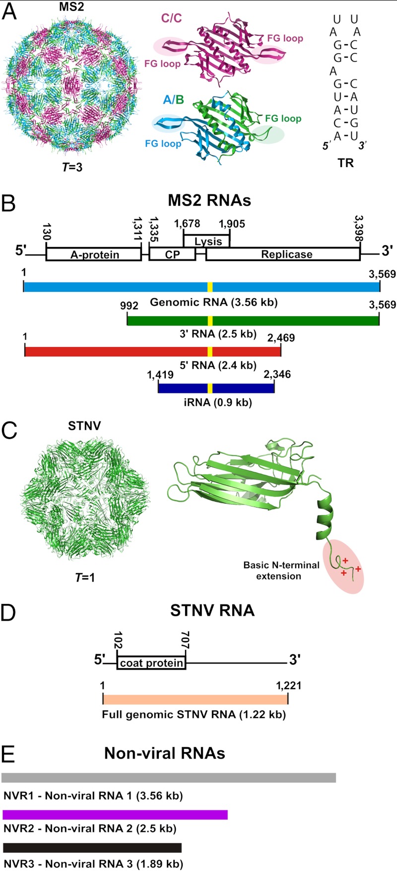
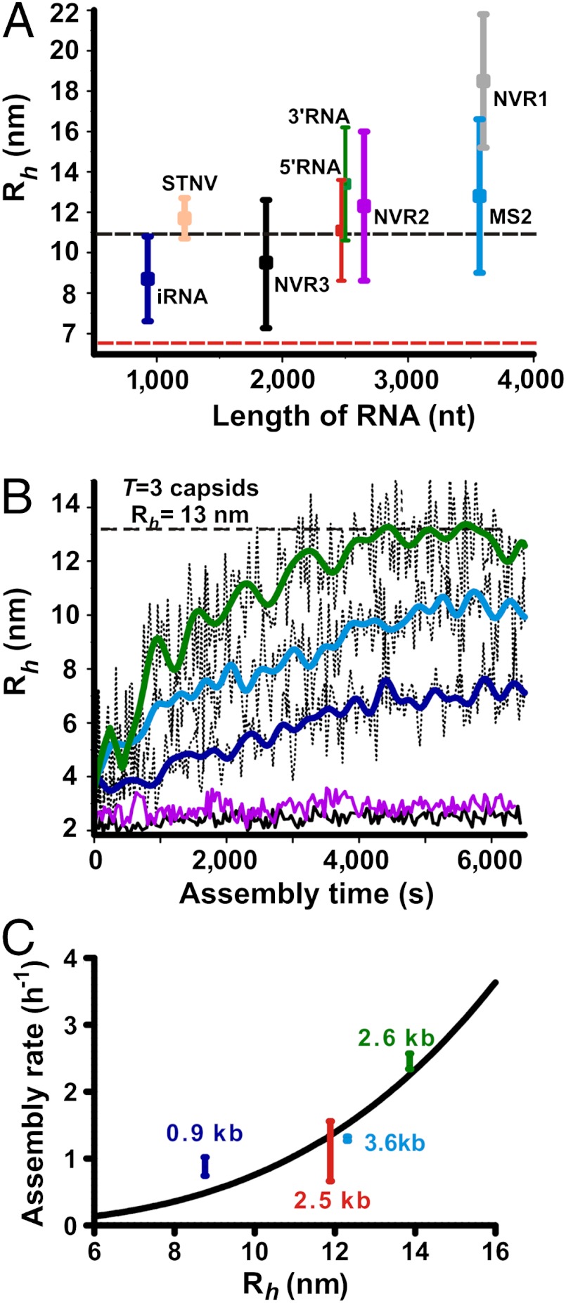
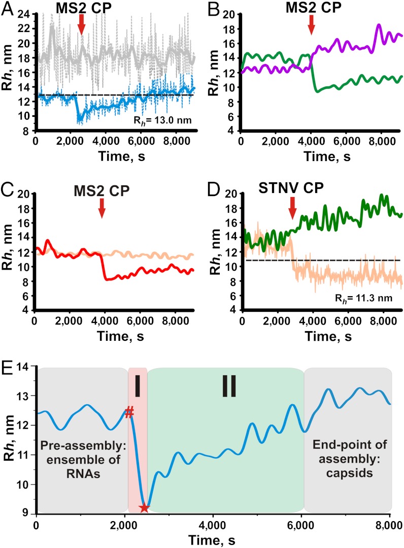
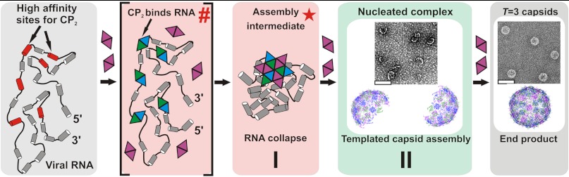
Genome packaging is an essential step in virus replication and a potential drug target. Single-stranded RNA viruses have been thought to encapsidate their genomes by gradual co-assembly with capsid subunits. In contrast, using a single molecule fluorescence assay to monitor RNA conformation and virus assembly in real time, with two viruses from differing structural families, we have discovered that packaging is a two-stage process. Initially, the genomic RNAs undergo rapid and dramatic (approximately 20-30%) collapse of their solution conformations upon addition of cognate coat proteins. The collapse occurs with a substoichiometric ratio of coat protein subunits and is followed by a gradual increase in particle size, consistent with the recruitment of additional subunits to complete a growing capsid. Equivalently sized nonviral RNAs, including high copy potential in vivo competitor mRNAs, do not collapse. They do support particle assembly, however, but yield many aberrant structures in contrast to viral RNAs that make only capsids of the correct size. The collapse is specific to viral RNA fragments, implying that it depends on a series of specific RNA-protein interactions. For bacteriophage MS2, we have shown that collapse is driven by subsequent protein-protein interactions, consistent with the RNA-protein contacts occurring in defined spatial locations. Conformational collapse appears to be a distinct feature of viral RNA that has evolved to facilitate assembly. Aspects of this process mimic those seen in ribosome assembly.


Conflict of interest statement

The authors declare no conflict of interest.

Figures

Fig. 1.
Fig. 1.
The bacteriophage MS2 and Satellite Tobacco Necrosis Virus (STNV) systems. (A) Structure of the T = 3 MS2 bacteriophage (PDB ID 2ms2). Structures of the A/B and C/C quasiequivalent coat protein dimers (PDB ID 1zdh) that differ in the conformations of their FG-loops (highlighted). Sequence and secondary structure of the high-affinity 19 nt TR stem-loop, which is known to cause a conformational switch in coat protein from the C/C to the A/B dimer. Capsid assembly is believed to be initiated at this site on genomic RNA. (B) Genetic map of the MS2 genome (GenBank Accession NC001417) and the RNAs used in assembly studies (color-coded here and throughout). The locations of TR are indicated by the yellow stripes. (C) Structure of the T = 1 STNV capsid. Structure of the STNV coat protein monomer (PDB ID 3RQV) shown with its positively charged N-terminal extension (highlighted). (D) Genetic map of the STNV genome (strain C, GenBank Accession AJ000898) and the RNA used in assembly studies. (E) Nonviral RNA controls used in the study (color-coded here and throughout).
Fig. 2.
Fig. 2.
(A) Size distribution of the test RNAs (color-coded as on Fig. 1) measured by FCS as a function of their lengths in nucleotides. Vertical error bars correspond to the full width at half maximum of Rh distributions for each of the RNAs. The black dashed line represents the inner capsid radius of MS2, whilst the red one corresponds to the inner radius of STNV capsid. (B) Assembly assays with fluorescently labeled MS2 CP2. Capsid assembly kinetics with genomic (blue), and subgenomic 3’RNA (green) and iRNA (dark blue). The kinetics with nonviral RNA controls are shown in magenta (NVR2) and black (NVR3). Apparent hydrodynamic radii, Rh, are plotted as a function of time from assembly initiation (assembly time). FFT smoothed data are shown as solid lines overlaid on the original traces (thin dotted lines). The gray dashed line represents the hydrodynamic radius of the MS2 virion. (C) Apparent assembly rates obtained from a single exponential approximation of Rh(t) for different MS2 RNAs as a function of their hydrodynamic size. Vertical error bars (rate) represent the standard error of repeated experiments.
Fig. 3.
Fig. 3.
Assembly with labelled long RNA was followed by Rh(t) and performed at 1∶200 molar ratio RNA∶CP2 for MS2 or at 1∶60 molar ratio for STNV under the conditions described in Materials and Methods . Data collection was started for RNA alone and the coat protein (CP) was mixed with RNA at the time points indicated by the red arrows. (A) gRNA (light blue) and NVR1 (gray). The black dashed line represents the Rh of the MS2 virion. (B) 3’RNA (green) and NVR2 (magenta). (C) 5’RNA (red) and STNV RNA (orange). (D) 3’RNA (green) & STNV RNA (orange) upon addition of STNV CP. The black dashed line represents the Rh of the STNV virion. (E) Changes in Rh for viral RNAs upon addition of cognate coat protein. Red hash (#) indicates the precollapse state of the RNA-protein complex, which is not detectable by FCS due to the fast kinetics of the CP binding reaction. The initial (ensemble of RNA molecules) and end-points of the assembly reaction (capsids) are highlighted in gray. The collapse stage (I) is highlighted in pink and the collapsed RNA intermediate is marked with a red star (*). The capsid assembly stage (II) is highlighted in green.
Fig. 4.
Fig. 4.
Proposed two-stage assembly model for MS2. A pre-assembly ensemble of protein-free genomic RNAs (gray box) presents a number of dispersed high affinity sites for CP2 (red, not to scale), having an Rh approximately 20–30% larger than the inner space of the T = 3 capsid. CP2 binds to these putative packaging signals creating an initiation complex denoted as a (hash) as in Fig. 3E. Protein–protein interactions within this complex trigger RNA collapse into the assembly intermediate with an Rh small enough to fit within the confines of the capsid shell, denoted by the (red star). This completes stage I of the assembly process (pink box). During stage II the stable collapsed complex recruits additional CP2 to populate shell-like species of the correct curvature (green box, see inset for EM) and allows efficient completion of capsids (end-point of assembly, gray box).

References

    1. Schneemann A. The structural and functional role of RNA in icosahedral virus assembly. Annu Rev Microbiol. 2006;60:51–67. - PubMed
    1. Lomonossoff GP. Pathogen-derived resistance to plant-viruses. Annu Rev Phytopathol. 1995;33:323–343. - PubMed
    1. Routh A, Domitrovic T, Johnson JE. Host RNAs, including transposons, are encapsidated by a eukaryotic single-stranded RNA virus. Proc Natl Acad Sci USA. 2012;109:1907–12. - PMC - PubMed
    1. Tumban E, Peabody J, Peabody DS, Chackerian B. A pan-HPV vaccine based on bacteriophage PP7 VLPs displaying broadly cross-neutralizing epitopes from the HPV minor capsid protein, L2. Plos One. 2011;6:e23310. - PMC - PubMed
    1. Simon AE, Gehrke L. RNA conformational changes in the life cycles of RNA viruses, viroids, and virus-associated RNAs. Biochim Biophys Acta. 2009;1789:571–583. - PMC - PubMed

Publication types

LinkOut - more resources