. 2012 Sep 25;109(39):15787-92.
doi: 10.1073/pnas.1204540109. Epub 2012 Sep 10.

TBC1D16 is a Rab4A GTPase activating protein that regulates receptor recycling and EGF receptor signaling

Affiliations

TBC1D16 is a Rab4A GTPase activating protein that regulates receptor recycling and EGF receptor signaling

Basem S Goueli et al. Proc Natl Acad Sci U S A. .

Abstract

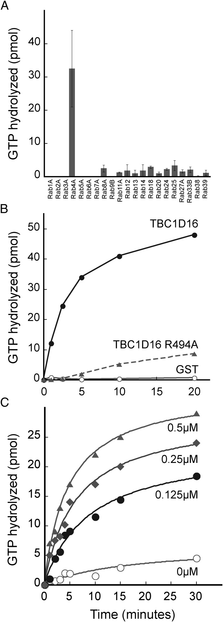
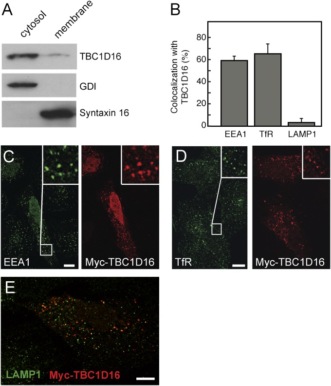
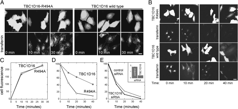
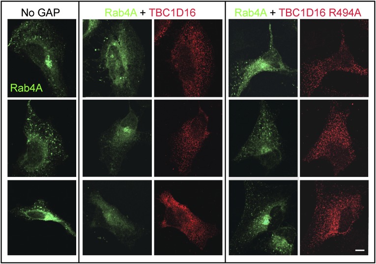
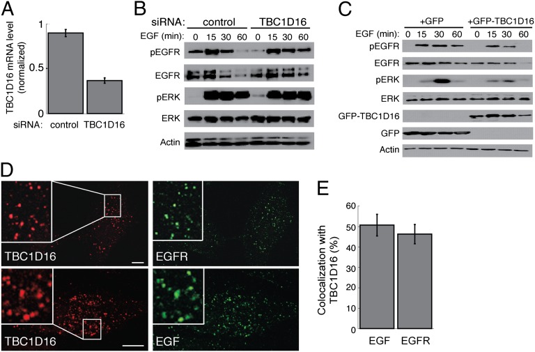
Rab4A is a master regulator of receptor recycling from endocytic compartments to the plasma membrane. The protein TBC1D16 is up-regulated in melanoma, and TBC1D16-overexpressing melanoma cells are dependent on TBC1D16. We show here that TBC1D16 enhances the intrinsic rate of GTP hydrolysis by Rab4A. TBC1D16 is both cytosolic and membrane associated; the membrane-associated pool colocalizes with transferrin and EGF receptors (EGFRs) and early endosome antigen 1, but not with LAMP1 protein. Expression of two TBC1D16 isoforms, but not the inactive R494A mutant, reduces transferrin receptor recycling but has no effect on transferrin receptor internalization. Expression of TBC1D16 alters GFP-Rab4A membrane localization. In HeLa cells, overexpression of TBC1D16 enhances EGF-stimulated EGFR degradation, concomitant with decreased EGFR levels and signaling. Thus, TBC1D16 is a GTPase activating protein for Rab4A that regulates transferrin receptor recycling and EGFR trafficking and signaling.


Conflict of interest statement

The authors declare no conflict of interest.

Figures

Fig. 1.
Fig. 1.
TBC1D16 is a GAP for Rab4A in vitro. (A) Biochemical screen of 21 Rabs. GAP activity was assayed using 150 pmol Rab loaded with γ-P32-GTP and 150 pmol GAP or GST control for 5 min at 25 °C. Depicted is the level of GAP-dependent, GTP hydrolysis observed. Error bars represent SD of duplicate determinations. (B) Reactions contained 150 pmol TBC1D16 (filled circles), TBC1D16 R494A (triangles), or GST (open circles) for the indicated times and 150 pmol Rab4A loaded with γ-P32-GTP. (C) Reactions were carried out as in B using TBC1D16 at the indicated concentrations with 1 μM Rab4A protein.
Fig. 2.
Fig. 2.
TBC1D16 colocalizes with transferrin receptor and EEA1. (A) Immunoblot of GFP-TBC1D16 comparing membrane and cytosol fractions. Control proteins were GDI for cytosol and Syntaxin 16 for membranes. Equal proportions of cytosol and membrane fractions were compared. (B) Quantitation of colocalization of TBC1D16 with EEA1 (471 TBC1D16+ structures in 10 cells), transferrin receptor (TfR) (646 structures as for EEA1), and LAMP1 (582 structures as for EEA1). (C–E) Immunofluorescence microscopy of HeLa cells expressing Myc-TBC1D16 and costained for EEA1 (C), TfR (D), or LAMP1 (E). In D, HeLa cells were serum-starved and then allowed to endocytose human holotransferrin for 30 min before being processed. All cells were subjected to freeze-thaw in liquid nitrogen to deplete cells of cytosolic proteins. The insets at the upper right of C and D are magnified portions (boxes) of the images shown in C and D. A merged image is shown in E. (Scale bars: 10 μm.)
Fig. 3.
Fig. 3.
TBC1D16 has no effect on transferrin internalization, but blocks transferrin recycling. (A) Cells transfected for 24 h with TBC1D16-R494A (Left) or WT TBC1D16 (Right) were incubated with rhodamine transferrin for the times indicated and then fixed for visualization. (B) Cells transfected as in A were pulsed with rhodamine transferrin for 30 min and then chased with unlabeled holotransferrin for the designated times to detect loss of internalized transferrin by recycling. (C) Quantitation of images obtained in an experiment carried out as in A. (D) Quantitation of images obtained in an experiment as in B. All experiments were performed three times; a representative example is shown. Each experiment involved counting 20 cells for each condition with <5% error observed at all time points. (Scale bars: 20 μm.) (E) UAC1273 cells at 48 h after transfection with control or fluorescent TBC1D16-siRNA were treated as in B. (Inset) Quantitative RT-PCR was used to determine relative TBC1D16 mRNA expression normalized to 18 S rRNA. The mean fluorescence per cell is shown. At least 80 cells were analyzed in each condition; only cells that incorporated fluorescent TBC1D16 siRNA were scored. Less than 5% error was observed at all time points.
Fig. 4.
Fig. 4.
TBC1D16 expression alters the appearance of GFP-Rab4A in HeLa cells. HeLa cells were transfected with GFP-Rab4A alone (Left), GFP-Rab4A with WT Myc-TBC1D16 (Middle), or Myc-TBC1D16-R494A (Right). GAP proteins were detected using anti-Myc antibodies (red) after cytosolic depletion using liquid nitrogen. (Scale bar: 10 μm.)
Fig. 5.
Fig. 5.
TBC1D16 regulates EGFR degradation, activation, and signaling. (A) Quantitative RT-PCR of TBC1D16 mRNA expression, normalized to 18S rRNA, in WM115 cells at 48 h after nucleofection with control or TBC1D16-siRNA. (B) Determination of phospho-EGFR (Y1068), EGFR, phospho-ERK (T183), ERK 1/2, and actin levels in WM115 cells (25 μg of total cell lysate) treated with 20 ng/mL EGF for indicated times at 48 h after nucleofection with control or TBC1D16 siRNA. Cells were serum-starved for 8 h and incubated with cycloheximide during the last 4 h before EGF stimulation. (C) HeLa cells (40 μg total lysate) at 24 h posttransfection with GFP or GFP-TBC1D16, analyzed as in B. (D) Colocalization of Myc-TBC1D16 with EGF and EGFR. At 24 h after transfection with Myc-TBC1D16, HeLa cells were were serum-starved for 30 min and then treated for 15 min with 100 ng/mL of Alexa Fluor 488 EGF for colocalization with EGF or 100 ng/mL of unlabeled human EGF for colocalization with EGFR. Insets show magnification of the images. (Scale bars: 10 μm.) (E) Quantitation of colocalization observed between EGF (618 TBC1D16+ structures) and EGFR (463 TBC1D16+ structures) with TBC1D16. The average colocalization for 10 cells is depicted.

References

    1. Stenmark H. Rab GTPases as coordinators of vesicle traffic. Nat Rev Mol Cell Biol. 2009;10:513–525. - PubMed
    1. Nguyen UT, et al. Analysis of the eukaryotic prenylome by isoprenoid affinity tagging. Nat Chem Biol. 2009;5:227–235. - PubMed
    1. Barr F, Lambright DG. Rab GEFs and GAPs. Curr Opin Cell Biol. 2010;22:461–470. - PMC - PubMed
    1. Fukuda M. TBC proteins: GAPs for mammalian small GTPase Rab? Biosci Rep. 2011;31:159–168. - PubMed
    1. Pan X, Eathiraj S, Munson M, Lambright DG. TBC-domain GAPs for Rab GTPases accelerate GTP hydrolysis by a dual-finger mechanism. Nature. 2006;442:303–306. - PubMed

Publication types

MeSH terms