. 2012 Sep 28:9:228.
doi: 10.1186/1742-2094-9-228.

Intravenous multipotent adult progenitor cell therapy after traumatic brain injury: modulation of the resident microglia population

Affiliations

Intravenous multipotent adult progenitor cell therapy after traumatic brain injury: modulation of the resident microglia population

Peter A Walker et al. J Neuroinflammation. .

Abstract

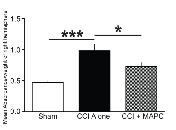
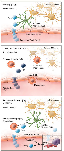
Introduction: We have demonstrated previously that the intravenous delivery of multipotent adult progenitor cells (MAPC) after traumatic brain injury affords neuroprotection via interaction with splenocytes, leading to an increase in systemic anti-inflammatory cytokines. We hypothesize that the observed modulation of the systemic inflammatory milieu is related to T regulatory cells and a subsequent increase in the locoregional neuroprotective M2 macrophage population.

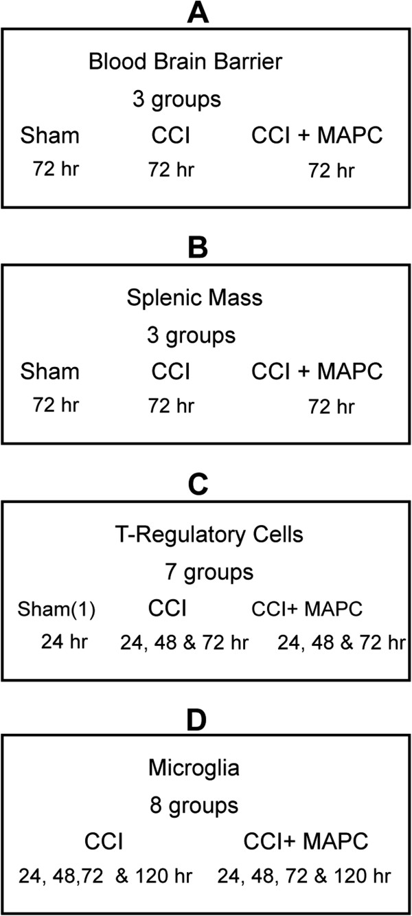
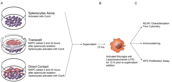
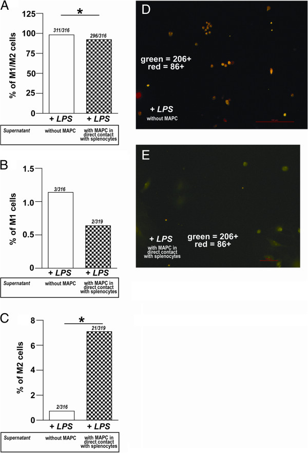
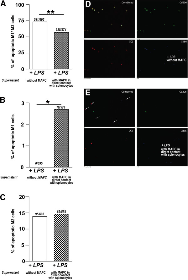
Methods: C57B6 mice were injected with intravenous MAPC 2 and 24 hours after controlled cortical impact injury. Animals were euthanized 24, 48, 72, and 120 hours after injury. In vivo, the proportion of CD4(+)/CD25(+)/FOXP3(+) T-regulatory cells were measured in the splenocyte population and plasma. In addition, the brain CD86(+) M1 and CD206(+) M2 macrophage populations were quantified. A series of in vitro co-cultures were completed to investigate the need for direct MAPC:splenocyte contact as well as the effect of MAPC therapy on M1 and M2 macrophage subtype apoptosis and proliferation.

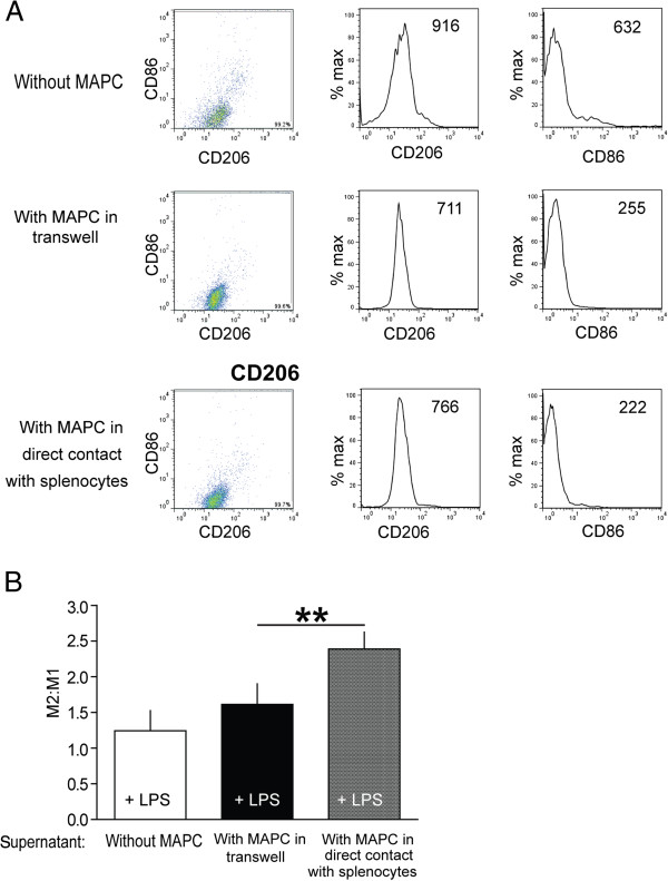
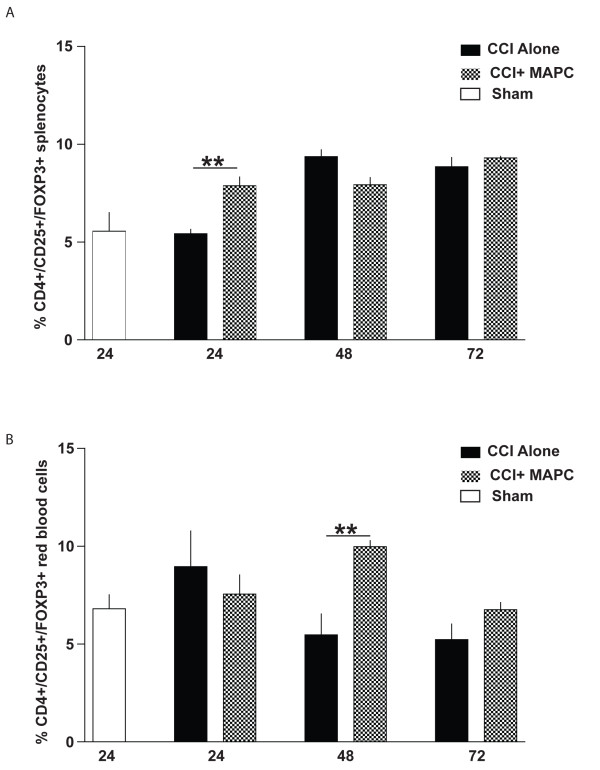
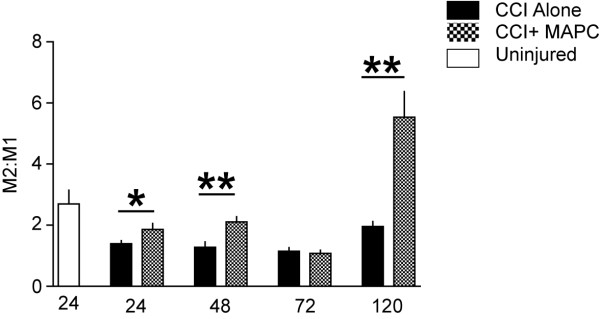
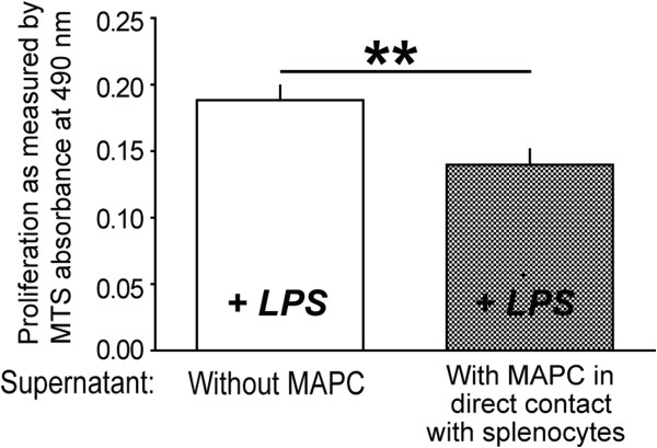
Results: Significant increases in the splenocyte and plasma T regulatory cell populations were observed with MAPC therapy at 24 and 48 hours, respectively. In addition, MAPC therapy was associated with an increase in the brain M2/M1 macrophage ratio at 24, 48 and 120 hours after cortical injury. In vitro cultures of activated microglia with supernatant derived from MAPC:splenocyte co-cultures also demonstrated an increase in the M2/M1 ratio. The observed changes were secondary to an increase in M1 macrophage apoptosis.

Conclusions: The data show that the intravenous delivery of MAPC after cortical injury results in increases in T regulatory cells in splenocytes and plasma with a concordant increase in the locoregional M2/M1 macrophage ratio. Direct contact between the MAPC and splenocytes is required to modulate activated microglia, adding further evidence to the central role of the spleen in MAPC-mediated neuroprotection.


Figures

Figure 1
Figure 1
Experimental design for in vivo experiments. Four sets of experiments were done in order to determine (A) blood brain barrier, (B) splenic mass, (C) characterization of T-regulatory cells in spleen and blood, and (D) characterization of local microglia phenotype using flow cytometry.
Figure 2
Figure 2
Schematic setup of in vitro MAPC, splenocyte, and microglia/macrophage interactions. (A) Isolated splenocytes are activated with 2 μg/ml of ConA. Two hours after isolation, MAPC were added at a ratio of 1:5 (MAPC:splenocyte) in co-culture or transwell cultures. 24 hours after the first dose of MAPCs, a second dose of MAPCs were added at the same ratio. 48 hours after the splenocyte harvest, supernatants were collected from each specific condition. (B) The supernatant was then used to stimulate LPS-activated microglia. (C) Microglia/macrophages were characterized via flow cytometry, immunostained or measured for proliferation using the MTS proliferation assay. LPS, lipopolysaccharide; MAPCs, multipotent adult progenitor cells; MTS.
Figure 3
Figure 3
Effect of MAPC:Splenocyte co-culture supernatant on stimulated microglial immunophenotype. (A) Samples of microglial cultures were first gated on forward- and side-scatter characteristics to exclude debris, electronic noise, and aggregates (not shown). Resulting populations were then gated to exclude a small number of artifacts displaying extremely high signal for either CD206-AF488 or CD86-PE. Rather than divide this microglia population further by imposing strict boundaries on a continuous expression pattern, mean fluorescence intensity (MFI, listed in each box) of the entire remaining population was then determined for each marker and the CD206/CD86 MFI ratio (M2:M1 ratio) calculated. (B) There was a significant increase in the M2:M1 ratio when the microglia received the supernatant derived from MAPC:splenocyte co-culture plus LPS when compared to microglia which only received LPS alone, or supernatant derived from MAPC:splenocyte transwell cultures plus LPS. ** represents P <0.01. LPS, lipopolysaccharide; MAPC, multipotent adult progenitor cell.
Figure 4
Figure 4
MAPC treatment reduces BBB permeability after TBI. BBB permeability measurement was completed using Evan’s blue dye as mean absorbance (nm) normalized to tissue weight (grams) derived from homogenized cortical tissue derived from the hemisphere ipsilateral to the CCI injury. Mice showed an increase in BBB permeability after CCI alone that was reversed by the intravenous injection of MAPC. * represents P <0.05 and *** represents P <0.005. BBB, blood brain barrier; MAPC, multipotent adult progenitor cell; TBI, traumatic brain injury.
Figure 5
Figure 5
MAPC treatment increases CD4+/CD25+/FOXP3+ cells after TBI in spleen and blood. (A) Measurement of CD4+/CD25+/FOXP3+ cells was completed in the splenocytes at 24, 48, and 72 hours after CCI injury and CCI injury plus MAPC (n = 4/group). There was a significant increase in T regulatory cells as a percentage of CD4+ T helper cells within the spleens of MAPC treated mice at 24 hours (P ≤0.01), with no difference seen at 48 or 72 hours. (B) In peripheral blood, percent T regulatory cells from MAPC treated mice was significantly higher at 48 hours after CCI injury (P ≤0.01). Of note, there is an additional control of sham (uninjured: white bar) for baseline profile of splenocytes. * represents P <0.05 and ** represents P <0.01. CCI, controlled cortical impact; MAPC, multipotent adult progenitor cell; TBI, traumatic brain injury.
Figure 6
Figure 6
MAPC treatment changes macrophage/microglia phenotype after TBI. Graph of the ratio of anti-inflammatory microglia versus proinflammatory microglia as determined by flow cytometry. Measurement of brain CD86+ M1 and CD206+ M2 macrophages was completed after cortical injury in mice at 24, 48, 72, and 120 hours after CCI injury (n = 6/group). There were significant increases in the ratio of M2 versus M1 mean fluorescence intensity between the CCI plus MAPC versus CCI alone at 24 (P ≤0.05), 48 (P ≤0.01) and 120 hours (P ≤0.01) after injury* represents P <0.05 and ** represents P <0.01. CCI, controlled cortical impact; MAPC, multipotent adult progenitor cell; TBI, traumatic brain injury.
Figure 7
Figure 7
Supernatant derived from MAPC:splencocyte co-cultures attenuates LPS induced proliferation. There was a significant reduction in proliferation of microglia as determined by absorbance of MTS (P ≤0.01: n = 8) in the microglia treated with supernatant derived from MAPC:splenocyte co-culture when compared to splenocyte only culture. ** represents P <0.01. LPS, lipopolysaccharide; MAPC, multipotent adult progenitor cell; MTS.
Figure 8
Figure 8
Supernatant derived from MAPC:splencocyte co-cultures promotes anti-inflammatory phenotype of microglia/macrophages. (A) Graph of percentage of M1/M2 activated with LPS and incubated with MAPC:splenocyte co-culture supernatant versus splenocyte culture supernatant (without MAPC). A significant decrease in the double positive microglial cells (CD86+ and CD206+) is seen in the MAPC:splenocyte co-culture group (P ≤0.05). (B) Fewer CD86+ microglia were observed in the MAPC:splenocyte co-culture group, but this change was not significant. (C) Significantly more CD206+ cells were observed in the MAPC:splenocyte co-culture group as opposed to the splenocyte alone group (P ≤0.05). (D) Photomicrograph of isolated microglia/macrophages activated LPS and splenocyte alone (without MAPC) supernatant. Note the number of dual stained cells. (E) Photomicrograph of isolated microglia/macrophages activated LPS and incubated with MAPC:splenocyte co-culture supernatant treatment. Note the number of M2 cells (green). M2 cells are labeled green and M1 cells are labeled red. * represents P <0.05. LPS, lipopolysaccharide; MAPC, multipotent adult progenitor cell.
Figure 9
Figure 9
Supernatant derived from MAPC:splenocyte co-cultures promotes apoptosis of proinflammatory microglia/macrophages. (A) Triple positive microglia (CD86+/CD206+/CC3+) significantly decreased after treatment with supernatant derived from MAPC:splenocyte co-cultures (P ≤0.01). (B) A significant increase in M1 microglial apoptosis was measured by the number of CD86+/CC3+ microglia in microglia treated with supernatant derived from MAPC:splenocyte co-cultures (P ≤0.05). (C) No significant difference in the number of CD206+/CC3+ microglia between the two groups was observed. (D) Photomicrograph of isolated microglia/macrophages activated LPS and splenocyte alone (without MAPC) supernatant. Note the number of triple stained cells. (E) Photomicrograph of isolated microglia/macrophages activated LPS and incubated with MAPC in direct contact with splenocytes supernatant treatment. Note the number of M1/CC3 as indicated by arrows. M2 positive cells are labeled green, CC3 positive cells are labeled red, and M1 positive cells are labeled blue * and ** represents P <0.05. LPS, lipopolysaccharide; MAPCs, multipotent adult progenitor cells.
Figure 10
Figure 10
Model of MAPC interactions with splenocytes to modulate microglia/macrophage phenotype. (A) In a normal uninjured brain resident microglia generally exist in a ramified M2 state. Functions include patrolling the brain to detect any disturbances or foreign substances as well as many other functions. This allows for a neuroprotection of neurons and other cell types. Regulatory T-cells (Treg) are in constant communication with the resident ramified microglia (B) After TBI, ramified microglia change into activated microglia, which function mainly to phagocytose cellular debris created after injury. In addition to the activated resident microglia, there is influx of macropahges due to the damaged (leaky) BBB as well as upregulation of chemoattractant molecules and adhesion factors. After injury activated microglia and infiltrating macrophages are indistinguishable from each other. Both these cell types are being modulated by effector T-cells (Teff). (C) After TBI, MAPC treatment helps seal the leaky BBB. In addition, with direct contact with splenocytes, MAPC treatment results in Treg proliferation. This in turn aids in modulating the activated microglia/macrophage into ramified microglia, thereby reducing the long-term proinflammatory effects of activated microglia. BBB, blood brain barrier; MAPC, multipotent adult progenitor cell; TBI, traumatic brain injury.

References

    1. Thurman DJ, Alverson C, Dunn KA, Guerrero J, Sniezek JE. Traumatic brain injury in the United States: A public health perspective. J Head Trauma Rehabil. 1999;14:602–615. doi: 10.1097/00001199-199912000-00009. - DOI - PubMed
    1. Olson JK, Miller SD. Microglia initiate central nervous system innate and adaptive immune responses through multiple TLRs. J Immunol. 2004;173:3916–3924. - PubMed
    1. Chirumamilla S, Sun D, Bullock MR, Colello RJ. Traumatic brain injury induced cell proliferation in the adult mammalian central nervous system. J Neurotrauma. 2002;19:693–703. doi: 10.1089/08977150260139084. - DOI - PubMed
    1. Beck KD, Nguyen HX, Galvan MD, Salazar DL, Woodruff TM, Anderson AJ. Quantitative analysis of cellular inflammation after traumatic spinal cord injury: evidence for a multiphasic inflammatory response in the acute to chronic environment. Brain. 2010;133:433–447. doi: 10.1093/brain/awp322. - DOI - PMC - PubMed
    1. Smith HS. Activated microglia in nociception. Pain Physician. 2010;13:295–304. - PubMed

Publication types

MeSH terms