. 2012 Oct 1;3(1):24.
doi: 10.1186/2041-9139-3-24.

Developmental and evolutionary origins of the pharyngeal apparatus

Affiliations

Developmental and evolutionary origins of the pharyngeal apparatus

Anthony Graham et al. Evodevo. .

Abstract

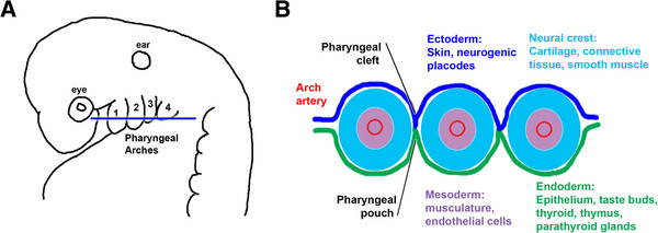
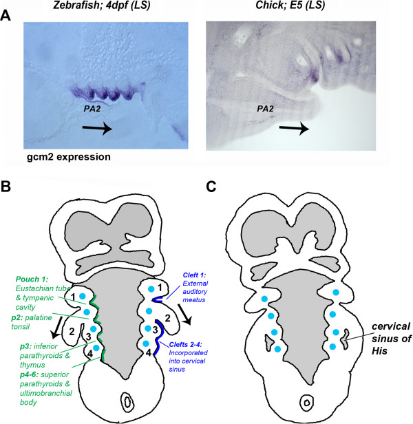
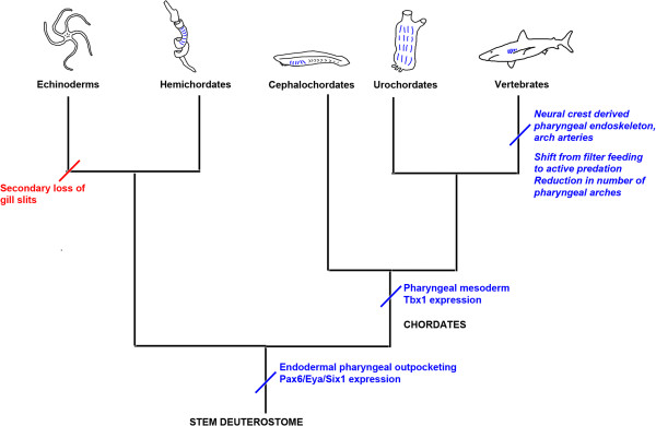
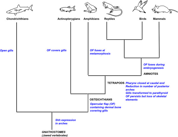
The vertebrate pharyngeal apparatus, serving the dual functions of feeding and respiration, has its embryonic origin in a series of bulges found on the lateral surface of the head, the pharyngeal arches. Developmental studies have been able to discern how these structures are constructed and this has opened the way for an analysis of how the pharyngeal apparatus was assembled and modified during evolution. For many years, the role of the neural crest in organizing pharyngeal development was emphasized and, as this was believed to be a uniquely vertebrate cell type, it was suggested that the development of the pharyngeal apparatus of vertebrates was distinct from that of other chordates. However, it has now been established that a key event in vertebrate pharyngeal development is the outpocketing of the endoderm to form the pharyngeal pouches. Significantly, outpocketing of the pharyngeal endoderm is a basal deuterostome character and the regulatory network that mediates this process is conserved. Thus, the framework around which the vertebrate pharyngeal apparatus is built is ancient. The pharyngeal arches of vertebrates are, however, more complex and this can be ascribed to these structures being populated by neural crest cells, which form the skeletal support of the pharynx, and mesoderm, which will give rise to the musculature and the arch arteries. Within the vertebrates, as development progresses beyond the phylotypic stage, the pharyngeal apparatus has also been extensively remodelled and this has seemingly involved radical alterations to the developmental programme. Recent studies, however, have shown that these alterations were not as dramatic as previously believed. Thus, while the evolution of amniotes was believed to have involved the loss of gills and their covering, the operculum, it is now apparent that neither of these structures was completely lost. Rather, the gills were transformed into the parathyroid glands and the operculum still exists as an embryonic entity and is still required for the internalization of the posterior pharyngeal arches. Thus, the key steps in our phylogenetic history are laid out during the development of our pharyngeal apparatus.


Figures

Figure 1
Figure 1
The vertebrate pharyngeal arches and their derivatives. (A) Lateral view of an amniote embryo, showing the characteristic bulges of the pharyngeal arches number 1 to 4 from anterior. The pouches intercalate between the arches. The position of the eye and ear are shown. (B) Schematic of a transverse section through the arch region, showing the constituent tissues: ectoderm, dark blue; endoderm, green; neural crest, pale blue ; mesoderm, purple.
Figure 2
Figure 2
Internalization of the posterior pharyngeal arches in amniotes. (A) Expression of gcm2 in zebrafish and chicks at comparable stages. In fish, this transcription factor is expressed in the pharyngeal pouches and their derivatives, the gill buds, and is required for their development. In chicks, gcm2 is also expressed in the pharyngeal pouches, which subsequently give rise to the parathyroids (modified from [3]). (B) Schematic of a transverse section through a human embryo, showing the second arch growing caudally over the posterior pouches (arrows). Internally, the pouches form several structures (indicated in green) derived from the endoderm. Ectodermal derivatives are indicated in dark blue. The posterior end of the pharynx is closed. (C) Later in development, the expanding second arch fuses with the body wall, enclosing the posterior arches and pouches. A sinus is formed, which is later obliterated. (B and C) adapted from Larsen [2].
Figure 3
Figure 3
Deuterostome phylogeny and the origin of pharyngeal segmentation. Acquisition of characteristics is indicated in blue, loss in red. The proposed stem deuterostome is likely to have possessed pharyngeal slits. These have been secondarily lost in echinoderms but retained in the hemichordates. Within the chordate lineage, cephalochordates (for example, amphioxus) and urochordates retain pharyngeal slits in the adult form (indicated by blue lines). Within the vertebrate lineage, there was a shift away from filter feeding towards active predation. Modification to the pharyngeal segments included a reduction in number, a neural-crest-derived endoskeletal support, and arch arteries providing vasculature for the gills.
Figure 4
Figure 4
Modification to the pharyngeal apparatus within the vertebrates. Within the vertebrates, the pharyngeal region has undergone extensive modification. The chondrichthyans retain open gill slits, but in the actinopterygian fish, these are covered by a bony operculum, which is derived from the second arch. The tetrapods have undergone the most radical remodelling of the pharyngeal arches as part of their adaptation to terrestrial life. Within this grouping, amphibians possess an opercular flap that fuses at metamorphosis; in amniote embryos, the second arch still expands caudally to cover the posterior arches, but does not retain skeletal elements, and later fuses to the cardiac eminence. The internal gill buds have become modified to form the parathyroid gland.

References

    1. Graham A, Smith A. Patterning the pharyngeal arches. Bioessays. 2001;23(1):54–61. doi: 10.1002/1521-1878(200101)23:1<54::AID-BIES1007>3.3.CO;2-X. - DOI - PubMed
    1. Schoenwolf GC, Bleyl SB, Brauer PR, Francis-West PH. Larsen's Human Embryology. Philadelphia: Churchill Livingstone; 2009.
    1. Richardson J, Shono T, Okabe M, Graham A. The presence of an embryonic opercular flap in amniotes. Proc Biol Sci. 2012;279(1727):224–229. doi: 10.1098/rspb.2011.0740. - DOI - PMC - PubMed
    1. Hoerstadius S, Sellman S. Experimentelle Untersuchungen ueber die Determination des Knorpeligen Kopfskelettes bei Urodelen. Nova Acta Regiae Soc Sci Ups Ser IV. 1946;13:1–170.
    1. Wagner G. Die Bedeutung der Neuralleiste fur die Kopfgestaltung der Amphibienlarven. Rev Suisse Zool. 1949;56:519–620.

LinkOut - more resources