Review
. 2012 Oct;22(5):651-9.
doi: 10.1016/j.sbi.2012.08.006. Epub 2012 Sep 25.

Time-resolved structural studies at synchrotrons and X-ray free electron lasers: opportunities and challenges

Affiliations
Review

Time-resolved structural studies at synchrotrons and X-ray free electron lasers: opportunities and challenges

Richard Neutze et al. Curr Opin Struct Biol. 2012 Oct.

Abstract

X-ray free electron lasers (XFELs) are potentially revolutionary X-ray sources because of their very short pulse duration, extreme peak brilliance and high spatial coherence, features that distinguish them from today's synchrotron sources. We review recent time-resolved Laue diffraction and time-resolved wide angle X-ray scattering (WAXS) studies at synchrotron sources, and initial static studies at XFELs. XFELs have the potential to transform the field of time-resolved structural biology, yet many challenges arise in devising and adapting hardware, experimental design and data analysis strategies to exploit their unusual properties. Despite these challenges, we are confident that XFEL sources are poised to shed new light on ultrafast protein reaction dynamics.


Figures

Figure 1
Figure 1
Time resolved Laue diffraction and WAXS studies of homodimeric hemoglobin (HbI). (A) Light minus dark 1.6 Å resolution Fobs-Fobs difference Fourier map of subunit B 100 ps after photodissociation of CO (green positive difference density contoured at 7σ; red negative difference density contoured at −7σ; σ is the rmsd electron density in the unit cell). Reproduced with permission from reference [21]. (B) Time-resolved difference WAXS curves ΔS(q,Δt) (laser on minus laser off; time-delay Δt indicated) recorded from HbI. Black curves, experimental data; red curves, modeled curves generated from linear combinations of three time-independent species-associated difference scattering curves derived from a kinetic analysis. (D) Population changes with time of the three intermediates I1, I2 and I3, for wild type HbI. (E) Schematic of the structural transitions for wild type HbI between the CO-bound form denoted HbI(CO)2 and the partly-liganded intermediates I1, I2 and I3. The green and blue arrows indicate the magnitudes and directions of the differences in heme-heme distances and subunit rotation angles relative to HbI(CO)2. Panels B, C, D and E are adapted with permission from [36].
Figure 2
Figure 2
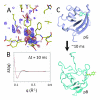
Time resolved Laue diffraction and WAXS studies of photoactive yellow protein (PYP). (A) Light minus dark 1.8 Å resolution Fobs-Fobs difference Fourier map 200 ns after photoactivation by light (blue/cyan positive difference density contoured at +3/+5 σ respectively; pink/red negative difference density contoured at −3/−4 σ respectively). The PYP pR intermediate is fully developed by 200 ns and decays on a time scale of ~100 μs. (B) Time resolved SAXS/WAXS data collected from PYP 10 ms after photoactivation. Black, experimental data; Red, theoretical model fit that incorporated additional information from other biophysical studies (static crystallography, DEER and NMR experiments). (C) Structural changes associated with the pG (resting) to pB conformations of PYP associated with the optimal fit to WAXS difference data shown in B. Panel A shows unpublished data provided by Marius Schmidt. Panels B and C are adapted with permission from reference [40].
Figure 3
Figure 3
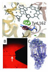
Time-resolved Laue diffraction and static serial femtosecond crystallography studies of the photosynthetic reaction centre from Bl. viridis (RCvir). (A) Light minus dark 2.95 Å Fobs-Fobs difference Fourier electron density observed 3 ms following photoactivation (green positive density contoured at 4 σ; red negative difference density contoured at −4 σ). A light induced movement of TyrL162 towards the special pair (P960) was modeled (grey resting conformation; blue photo-activated conformation) due to P960 being photo-oxidized. (B) Microjet used to inject lipidic sponge phase samples containing microcrystals of RCvir. The jet is approximately 4 μm across where the XFEL beam interacts with the jet (white arrow). (C) 2Fobs-Fcalc electron density map (contoured at 1 σ) recovered from the serial femtosecond crystallography (SFX) structure of RCvir at 8.2 Å resolution [51]. Panel A is adapted with permission from reference [23]. Panels B and C are reproduced with permission from reference [51].

References

    1. Ihee H, Wulff M, Kim J, S. I. A: Ultrafast X-ray scattering: structural dynamics from diatomic to protein molecules. Int Rev Phys Chem. 2010;29:453–520.
    1. Cammarata M, Eybert L, Ewald F, Reichenbach W, Wulff M, Anfinrud P, Schotte F, Plech A, Kong Q, Lorenc M, Lindenau B, et al. Chopper system for time resolved experiments with synchrotron radiation. Rev Sci Instrum. 2009;80:015101. - PubMed
    1. Schotte F, Lim M, Jackson TA, Smirnov AV, Soman J, Olson JS, Phillips GN, Jr., Wulff M, Anfinrud PA. Watching a protein as it functions with 150-ps time-resolved x-ray crystallography. Science. 2003;300:1944–1947. - PubMed
    1. Cho HS, Dashdorj N, Schotte F, Graber T, Henning R, Anfinrud P. Protein structural dynamics in solution unveiled via 100-ps time-resolved x-ray scattering. Proc Natl Acad Sci U S A. 2010;107:7281–7286. * This paper provides tantalizing clues that significant conformational changes are occurring in Mb:CO on a time scale faster than 100 ps, which is the current state-of-the-art in time resolved WAXS at synchrotron sources.

    1. Emma P, Akre R, Arthur J, Bionta R, Bostedt C, Bozek J, Brachmann A, Bucksbaum PH, Coffee R, Decker FJ, Ding Y, et al. First lasing and operation of an angstrom-wavelength free-electron laser. Nat Photon. 2010;4:641–647. * This is a landmark paper marking the first lasing at hard X-ray wavelengths at the LCLS.

Publication types

LinkOut - more resources