. 2012 Sep 28;151(1):167-80.
doi: 10.1016/j.cell.2012.07.034.

Dual binding of chromomethylase domains to H3K9me2-containing nucleosomes directs DNA methylation in plants

Affiliations

Dual binding of chromomethylase domains to H3K9me2-containing nucleosomes directs DNA methylation in plants

Jiamu Du et al. Cell. .

Abstract

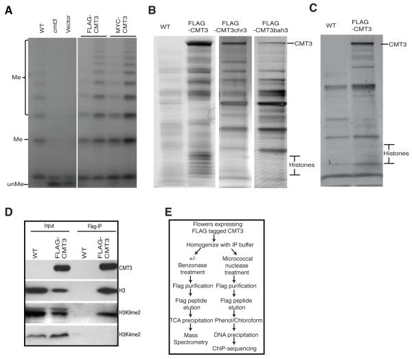
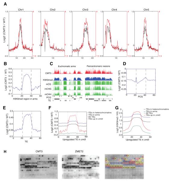
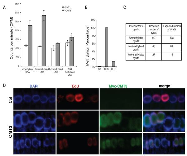
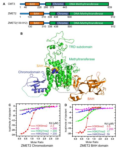
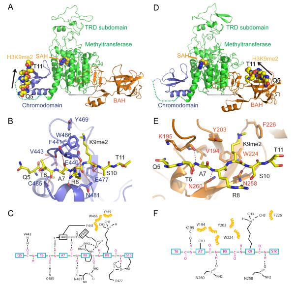
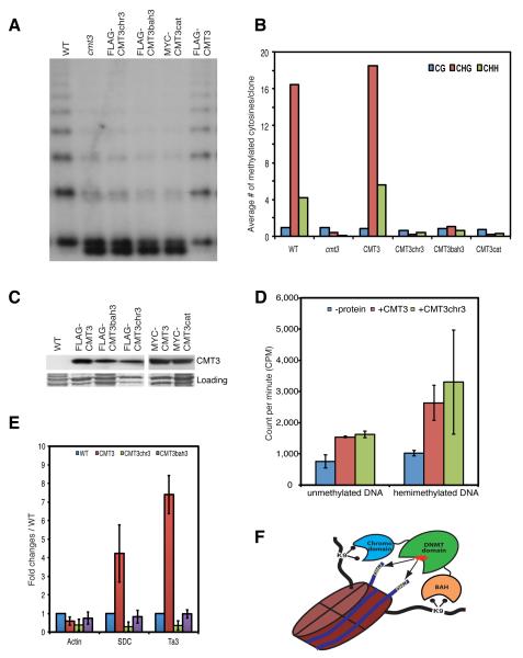
DNA methylation and histone modification exert epigenetic control over gene expression. CHG methylation by CHROMOMETHYLASE3 (CMT3) depends on histone H3K9 dimethylation (H3K9me2), but the mechanism underlying this relationship is poorly understood. Here, we report multiple lines of evidence that CMT3 interacts with H3K9me2-containing nucleosomes. CMT3 genome locations nearly perfectly correlated with H3K9me2, and CMT3 stably associated with H3K9me2-containing nucleosomes. Crystal structures of maize CMT3 homolog ZMET2, in complex with H3K9me2 peptides, showed that ZMET2 binds H3K9me2 via both bromo adjacent homology (BAH) and chromo domains. The structures reveal an aromatic cage within both BAH and chromo domains as interaction interfaces that capture H3K9me2. Mutations that abolish either interaction disrupt CMT3 binding to nucleosomes and show a complete loss of CMT3 activity in vivo. Our study establishes dual recognition of H3K9me2 marks by BAH and chromo domains and reveals a distinct mechanism of interplay between DNA methylation and histone modification.


Figures

Figure 1
Figure 1. CMT3 is Associated with Histones in vivo
(A) Complementation analysis by southern blot at 5S rDNA locus. Genomic DNA was digested with the methylation-sensitive MspI restriction enzyme. The (unMe) DNA fragment was the unmethylated DNA and (Me) DNA fragments were generated when MspI digestion was blocked by DNA methylation. The two lanes indicated by a horizontal line are from two independent transgenetic plant lines. Wild type Columbia ecotype (WT) is a control. (B) Silver stained SDS-PAGE gel showed the presence of histone proteins in CMT3, but not in CMT3chr3 and CMT3bah3. (C) Histone proteins were absent from protein complexes after benzonase treatment. (D) CMT3 specifically pulls down H3K9me2. (E) Outline of CMT3 affinity purification procedure for mass spectrometry and ChIP-seq. See also Tables S1-S2.
Figure 2
Figure 2. CMT3 Is Enriched in Heterochromatic Regions and Highly Correlated with H3K9me2
(A) Distribution of CMT3 (black) and H3K9me2 (red) along the five Arabidopsis chromosomes. (B) CMT3 ChIP-seq signal is enriched in heterochromatic patches in the arms. (C) Genome browser views of pericentromeric and euchromatic regions. mCG: CG methylation; mCHG: CHG methylation; mCHH: CHH methylation; TE: transposable element; PCG: protein coding gene. (D,E) CMT3 ChIP-seq signal is not enriched in the protein coding gene regions (D), but is enriched in TEs (E). (F,G) CMT3 (F) and H3K9me2 (G) are enriched in TEs that are upregulated in cmt3 null mutants. (H) CMT3 and ZMET2 specifically bind to mono-, di- and tri-methylation of H3K9. Blue, red and yellow circles in right panel represent the locations of mono-, di- and tri-methylation at lysine 9-containing peptides on the array, respectively. See also Figure S1A.
Figure 3
Figure 3. CMT3 Is Primarily a CHG Methyltransferase and Predominantly Expressed in Actively Replicating Cells
(A) CMT3 has activity on both unmethylated and hemi-methylated DNA oligos, but not fully methylated DNA oligos containing methylations at all cytosines. Error bars represent the standard deviation of three replicates. (B) Bisulfite sequencing of in vitro methylated plasmid DNA. (C) Hemimethylated dyads at the Ta3 locus were significantly lower than expected (Chi square value: 32.84). Probability of obtaining the observed distribution by random chance is less than 0.1%. (D) Accumulation of CMT3 in cells with active replication. DAPI staining shows the localization of nuclei (Blue). Actively replicating cells were labeled by 5-ethynul-2′-deoxyuridine (EdU, Red) and CMT3 was immuno-stained by MYC antibody (Green). See also Figures S1B and Tables S3 and S4.
Figure 4
Figure 4. Binding to Methylated Peptides and the Structure of ZMET2 in the Free State
(A) Color-coded domain architecture of full length CMT3, ZMET2, and the ZMET2 (130-912) construct used to grow crystals. (B) Ribbon representation of the structure of ZMET2 with bound SAH. The BAH, methyltransferase, and chromo domains are colored in orange, green, and blue, respectively, with the bound SAH molecule shown in a space filling representation. Some disordered regions were not built in the final model and are shown as dashed lines. (C, D) ITC binding curves for complex formation between ZMET2 chromo (C) and BAH (D) domains and H3K9me2, H3K27me2, H4K20me2, and unmodified H3 peptides. Kd values are listed as an insert. See also Figure S2 and Table S5.
Figure 5
Figure 5. Structure of Chromo and BAH Domains of ZMET2 with Bound H3K9me2 Peptide
(A, D) Ribbon representation of the structure of ZMET2(130-912) complexes with H3(1-15)K9me2 (A) and H3(1-32)K9me2 peptide (D). The color-coding is the same as in Figure 4B. The bound peptide and SAH are shown in a space-filling representation, with the orientation of the bound peptide shown by an arrow. (B, E) Intermolecular interactions between the chromodomain of ZMET2 and H3(1-15)K9me2 peptide (B) and between the BAH domain of ZMET2 and H3(1-32)K9me2 peptide (E). The dimethylammonium group of K9 is accommodated within an aromatic cage. Intermolecular hydrogen bonds are designated by dashed red lines. (C, F) A schematic representation of the intermolecular interactions between the chromodomain of ZMET2 and the bound H3(1-15)K9me2 peptide (C) and between the BAH domain of ZMET2 and the bound H3(1-32)K9me2 peptide (F). See also Figures S3, S4, and S5 and Table S5.
Figure 6
Figure 6. Both BAH and Chromo Domains Are Important for CMT3 Function in vivo.
(A) Southern blot of the 5S rDNA locus. (B) Graphical representation of bisulfite sequencing analysis of Ta3 locus. (C) Western blots showing the expression of the wild-type and mutant versions of the CMT3 protein. The bottom panel is the Coomassie blue staining and serves as a loading control. (D) In vitro activity assay. Error bars represent the standard deviation of three biological replicates. (E) ChIP data showing the failure of CMT3chr3 and CMT3bah3 to bind to chromatin at Ta3 and SDC. Error bars represent the standard deviation of three biological replicates. (F) A working model of dual recognition mechanism of CMT3 BAH and chromo domains simultaneously reading the H3K9me2 on a single nucleosome to position the methyltransferase domain over the nucleosomal DNA. See also Figures S6 and Table S2.

References

    1. Adams PD, Afonine PV, Bunkoczi G, Chen VB, Davis IW, Echols N, Headd JJ, Hung LW, Kapral GJ, Grosse-Kunstleve RW, et al. PHENIX: a comprehensive Python-based system for macromolecular structure solution. Acta Crystallogr D Biol Crystallogr. 2010;66:213–221. - PMC - PubMed
    1. Armache KJ, Garlick JD, Canzio D, Narlikar GJ, Kingston RE. Structural basis of silencing: Sir3 BAH domain in complex with a nucleosome at 3.0 A resolution. Science. 2011;334:977–982. - PMC - PubMed
    1. Bartee L, Bender J. Two Arabidopsis methylation-deficiency mutations confer only partial effects on a methylated endogenous gene family. Nucleic Acids Res. 2001;29:2127–2134. - PMC - PubMed
    1. Bernatavichute YV, Zhang X, Cokus S, Pellegrini M, Jacobsen SE. Genome- wide association of histone H3 lysine nine methylation with CHG DNA methylation in Arabidopsis thaliana. PLoS ONE. 2008;3:e3156. - PMC - PubMed
    1. Bestor TH, Ingram VM. Two DNA methyltransferases from murine erythroleukemia cells: purification, sequence specificity, and mode of interaction with DNA. Proc Natl Acad Sci U S A. 1983;80:5559–5563. - PMC - PubMed

Publication types