Review
. 2013 Mar;189(3):803-11.
doi: 10.1016/j.juro.2012.05.078. Epub 2012 Sep 25.

Reactive oxygen species as the molecular modulators of calcium oxalate kidney stone formation: evidence from clinical and experimental investigations

Affiliations
Review

Reactive oxygen species as the molecular modulators of calcium oxalate kidney stone formation: evidence from clinical and experimental investigations

Saeed R Khan. J Urol. 2013 Mar.

Abstract

Purpose: Idiopathic calcium oxalate kidney stones form while attached to Randall plaques, the subepithelial deposits on renal papillary surfaces. Plaque formation and growth mechanisms are poorly understood. Plaque formation elsewhere in the body is triggered by reactive oxygen species and oxidative stress. This review explores possible reactive oxygen species involvement in plaque formation and calcium oxalate nephrolithiasis.

Materials and methods: A search of various databases for the last 8 years identified literature on reactive oxygen species involvement in calcium oxalate nephrolithiasis. The literature was reviewed and results are discussed.

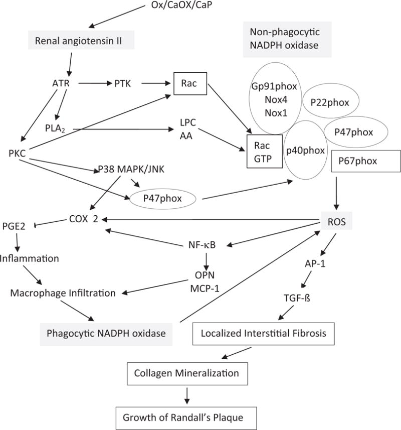
Results: Under normal conditions reactive oxygen species production is controlled, increasing as needed and regulating crystallization modulator production. Reactive oxygen species overproduction or decreased antioxidants lead to oxidative stress, inflammation and injury, and are involved in stone comorbidity. All major chronic inflammation markers are detectable in stone patient urine. Patients also have increased urinary excretion of the IαI and the thrombin protein families. Results of a recent study of 17,695 participants in NHANES III (National Health and Nutrition Examination Survey) showed significantly lower antioxidants, carotene and β-cryptoxanthin in those with a kidney stone history. Animal model and tissue culture studies revealed that high oxalate, calcium oxalate and calcium phosphate crystals provoked renal cell reactive oxygen species mediated inflammatory responses. Calcium oxalate crystals induce renin up-regulation and angiotensin II generation. Nonphagocytic NADPH oxidase leads to reactive oxygen species production mediated by protein kinase C. The P-38 MAPK/JNK transduction pathway is turned on. Transcriptional and growth factors, and generated secondary mediators become involved. Chemoattractant and osteopontin production is increased and macrophages infiltrate the renal interstitium around the crystal. Phagocytic NADPH oxidase is probably activated, producing additional reactive oxygen species. Localized inflammation, extracellular matrix and fibrosis develop. Crystallization modulators have a significant role in inflammation and tissue repair.

Conclusions: Based on available data, Randall plaque formation is similar to extracellular matrix mineralization at many body sites. Renal interstitial collagen becomes mineralized, assisting plaque growth through the interstitium until the mineralizing front reaches papillary surface epithelium. Plaque exposure to pelvic urine may also be a result of reactive oxygen species triggered epithelial sloughing.


Figures

Figure 1
Figure 1
Transmission electron microscopy. A, spherulitic CaP deposits in kidneys of idiopathic stone former in interstitium and around necrosing tubules (arrows). Scale bar indicates 10 μm. B, higher magnification reveals CaP (arrowhead) and collagen fibers (arrows). Bar indicates 1 μm.
Figure 2
Figure 2
Scanning electron microscopy shows NRK52E cells on exposure to 133 μg/cm2 hydroxyapatite. A, normal cells. B, higher magnification shows surface covered with short microvilli. C, crystals are evenly dispersed on cell surface. D, at 3 hours of treatment crystals (x) appear to be in contact with thin projections from cell surface. E, cell appears to take in crystals. Arrows indicate cell leading edge. F, epithelial cell appears to have phagocytosed crystals. Arrows indicate cell leading edge.
Figure 3
Figure 3
Light microscopy. A, rat kidney with significant CaOx crystal deposition as seen under polarized light. H & E, reduced from ×1.2. B, in kidney section crystals appear as dark deposits. Pizzalato stain, reduced from ×10. C, kidney of hyperoxaluric rat treated with apocynin demonstrates highly significant decrease in CaOx crystal deposition. Reduced from ×10.
Figure 4
Figure 4
Immunohistochemical staining of kidneys of hyperoxaluric rats. A, ED-1 positive cells were present in renal interstitium next to birefringent CaOx crystal containing tubules, indicating infiltration of monocytes and macrophages (inflammatory response). Reduced from ×20. B, tubular epithelial cells stained positive for CD-44, which is receptor for hyaluronic acid and other ligands, such as OPN and collagens. Reduced from ×20. C, renal papillary surface epithelium in renal fornix, particularly outer surface, shows heavy staining for OPN, modulator of biomineralization and inflammation. Reduced from ×40. D, tubular epithelial cells demonstrate positive staining for proliferating cell nuclear antigen, indicating cellular regeneration and proliferation. Reduced from ×20. E, increased staining for OPN in renal cortical tubules. Reduced from ×20. F, epithelial cells lining cortical renal tubules stained positive for E-cadherin, which has important role in cell adhesion. Reduced from ×20. G, tubular epithelial cells showed increased staining for NF-κB, mediator of inflammation and injury. H, crystal deposition was associated with increased expression of kidney injury molecule in tubular cells and their luminal surfaces. Reduced from ×20.
Figure 5
Figure 5
Pathways involved in NADPH oxidase regulation and ROS production. Renal cell exposure to high Ox, CaOx/CaP crystals and mechanical stress associated with crystal deposition in kidneys leads to renin up-regulation and angiotensin II generation. Nonphagocytic NADPH oxidase is activated and ROS are produced, mediated by PKC. Activation involves phosphorylation of p47phox, and translocation of Rac1 and p47phox to membrane. P-38 mitogen activated protein kinase/jun N-terminal kinase (MAPK/JNK) transduction pathway is turned on. Various transcriptional and growth factors, including NF-κB, AP-1 and transforming growth factor-β (TGF- β), become involved. Secondary mediators are generated, such as isoprostanes, cytoplasmic phospholipase A2 and prostaglandin. Production of chemoattractants, such as MCP-1 and crystallization modulator OPN, is increased. Macrophages infiltrate renal interstitium around crystal deposits. Activation of phagocytic NADPH oxidase results in additional ROS production. Inflammation, fibrosis, collagen deposition and mineralization develop, leading to growth of interstitial CaP deposits or RP. AA, arachidonic acid. ATR, angiotensin II type 1 receptor. COX 2, cyclooxygenase 2. LPC, lysophosphatidylcholine. PGE2, prostaglandin E2. PLA2, phospholipase A2. PTK, protein tyrosine kinase. Rac GTP, ρ-related guanosine triphosphate.

References

    1. Brikowski TH, Lotan Y, Pearle MS. Climate-related increase in the prevalence of urolithiasis in the United States. Proc Natl Acad Sci U S A. 2008;105:9841. - PMC - PubMed
    1. McAteer JA, Evan AP. The acute and longterm adverse effects of shock wave lithotripsy. Semin Nephrol. 2008;28:200. - PMC - PubMed
    1. Coe FL, Evan AP, Lingeman JE, et al. Plaque and deposits in nine human stone diseases. Urol Res. 2010;38:239. - PMC - PubMed
    1. Khan SR. Hyperoxaluria-induced oxidative stress and antioxidants for renal protection. Urol Res. 2005;33:349. - PubMed
    1. Manea A. NADPH oxidase-derived reactive oxygen species: involvement in vascular physiology and pathology. Cell Tissue Res. 2010;342:325. - PubMed