. 2013 Feb:65:65-73.
doi: 10.1016/j.neuropharm.2012.09.015. Epub 2012 Sep 26.

Ciliary neurotrophic factor activates NF-κB to enhance mitochondrial bioenergetics and prevent neuropathy in sensory neurons of streptozotocin-induced diabetic rodents

Affiliations

Ciliary neurotrophic factor activates NF-κB to enhance mitochondrial bioenergetics and prevent neuropathy in sensory neurons of streptozotocin-induced diabetic rodents

Ali Saleh et al. Neuropharmacology. 2013 Feb.

Abstract

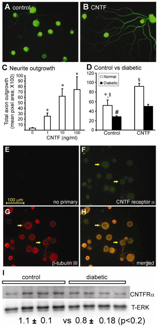
Diabetes causes mitochondrial dysfunction in sensory neurons that may contribute to peripheral neuropathy. Ciliary neurotrophic factor (CNTF) promotes sensory neuron survival and axon regeneration and prevents axonal dwindling, nerve conduction deficits and thermal hypoalgesia in diabetic rats. In this study, we tested the hypothesis that CNTF protects sensory neuron function during diabetes through normalization of impaired mitochondrial bioenergetics. In addition, we investigated whether the NF-κB signal transduction pathway was mobilized by CNTF. Neurite outgrowth of sensory neurons derived from streptozotocin (STZ)-induced diabetic rats was reduced compared to neurons from control rats and exposure to CNTF for 24 h enhanced neurite outgrowth. CNTF also activated NF-κB, as assessed by Western blotting for the NF-κB p50 subunit and reporter assays for NF-κB promoter activity. Conversely, blockade of NF-κB signaling using SN50 peptide inhibited CNTF-mediated neurite outgrowth. Studies in mice with STZ-induced diabetes demonstrated that systemic therapy with CNTF prevented functional indices of peripheral neuropathy along with deficiencies in dorsal root ganglion (DRG) NF-κB p50 expression and DNA binding activity. DRG neurons derived from STZ-diabetic mice also exhibited deficiencies in maximal oxygen consumption rate and associated spare respiratory capacity that were corrected by exposure to CNTF for 24 h in an NF-κB-dependent manner. We propose that the ability of CNTF to enhance axon regeneration and protect peripheral nerve from structural and functional indices of diabetic peripheral neuropathy is associated with targeting of mitochondrial function, in part via NF-κB activation, and improvement of cellular bioenergetics.


Figures

Fig. 1
Fig. 1
CNTF significantly increased axonal outgrowth in normal and, to a lesser extent, in diabetic neurons. Cultured adult DRG sensory neurons from normal rats were grown for 1 day in the absence of neurotrophic factors with a range of cytokines under defined conditions and then fixed and stained for neuron-specific β-tubulin III. In (A) is untreated, or (B) treated with CNTF (10 ng/ml). In (C) is shown total axon outgrowth of neurons in response to a range of CNTF concentrations. Values are means ± SEM (n=3 replicates). *p<0.05 vs control (oneway ANOVA). In (D) is shown total axon outgrowth for sensory neurons isolated from control (white column) or 3–5 month STZ-diabetic rats (black column) cultured for 1 day ± CNTF (at 10ng/ml). Values are the means ± SEM (n=3 replicates). For control, *p<0.05 vs CNTF; for diabetic, # p<0.05 vs CNTF, § p<0.05 vs diabetic. DRG sensory neurons from normal rats were cultured for 1 day in the absence of neurotrophic factors and were fixed and immunostained with (E) no antibody, (F) stained with CNTF receptor α antibody, (G) with β-tubulin III antibody, and (H) merge of F and G. The white arrows indicate co-localization of receptor and neuron-specific staining. The bar = 100μm. (I) Western blot for CNTF receptor α comparing control and diabetic cultures at 24 h in vitro with quantification. Expression levels are indicated below the blots and are expressed relative to T-ERK and are means ± SEM (n=4 replicates); no significant difference between groups.
Fig. 2
Fig. 2
Inhibition of NF-κB by SN50 impairs cytokine-mediated axon outgrowth. DRG sensory neurons from normal rats were cultured for 1 day in the presence of low dose neurotrophic factors (to improve viability in presence of SN50) and were exposed to the NF-κB peptide inhibitor SN50 (10 μg/ml), or its control peptide SN50M (10 μg/ml) ± CNTF (10ng/ml). Neurons were fixed and stained for neuron-specific β-tubulin III. Shown is (A) no treatment, (B) + SN50, (C) + CNTF, and (D) + CNTF + SN50. In (E) is shown the impact on total axon outgrowth. Data are mean ± SEM (n=3 replicates). *p<0.05 vs. control, or SN50, or SN50M; **p<0.05 vs. CNTF (by one way ANOVA).
Fig. 3
Fig. 3
CNTF activates NF-κB in adult DRG sensory neuron culture. Cultured DRG sensory neurons from normal rats, in the presence of low dose neurotrophic factors, were transfected with WT-NF-κB promoter. One day after transfection with functional WT-NF-κB promoter the neurons were stimulated for 12 h with a range of doses of CNTF (A). NF-κB-induced luciferase activity was normalized to Renilla luciferase activity. Fold induction represents luciferase activity in CNTF-treated cells compared with untreated cells and is the mean of three independent experiments. Values are means ± SEM, n=3; *p<0.05 vs control. In (B) neurons from control or STZ-induced diabetic rats were transfected with WT-NF-κB promoter and were then treated for 12 h with CNTF (10 ng/ml). Values are mean ± SEM, n=3; *p<0.05 vs. control. (C) shows basal NF-κB reporter activity in cultures from age matched control rats vs STZ-diabetic. Values are means ± SEM, n=3; *p<0.05 vs. control. In (D) are shown representative Western blots and quantification for NF-κB p50 and T-ERK showing effect of CNTF (10 ng/ml) treatment for up to 24 h on expression. Values are mean ± SEM, n=3, *p<0.05 vs control (by one way ANOVA).
Fig. 4
Fig. 4
Decreased NF-κB subunit p50 expression is normalized in DRG from STZ-diabetic mice treated with CNTF. Age matched control and STZ-diabetic C57Bl6/J mice were maintained for 18 wk and then a sub-group of diabetic mice treated systemically with 2 mg/kg CNTF thrice weekly for the final 12 wk. In (A) are shown Western blots of DRG homogenates probed for NF-κB p50 and total ERK (loading control; each lane represents an individual mouse) and (B) bar chart of quantification of p50 expression levels. In (C) is presented DNA binding activity of DRG homogenates. Values are means ± SEM, n=5; *p<0.05 vs control or Db + CNTF (one way ANOVA).
Fig. 5
Fig. 5
Mitochondrial bioenergetics is abnormal in cultured neurons from diabetic mice and is corrected by CNTF. OCR was measured at basal level with subsequent and sequential addition of oligomycin (1μM), FCCP (1.0 μM), and rotenone (1 μM) + antimycin A (AA; 1 μM) to DRG neurons cultured from age-matched control (blue) and 3–5 month STZ-induced diabetic mice (black) in the presence of low dose neurotrophic factors. Oligomycin inhibits the ATP synthase leading to a build-up of the proton gradient and dearth of ATP that inhibits electron flux and reveals the state of coupling efficiency. Uncoupling of the respiratory chain by FCCP injection reveals the maximal capacity to reduce oxygen. Finally, rotenone + antimycin A were injected to inhibit the flux of electrons through complexes I and III, and thus no oxygen was further consumed at cytochrome c oxidase. The remaining OCR determined after this intervention is primarily non-mitochondrial. Data are expressed as OCR in pmoles/min for 1,000 cells (there were approximately 2,500–5,000 cells per well). Dotted lines, a-e in (A) have been used later for quantification of bioenergetics parameters. The OCR measurements in (A) control (blue) and diabetic (black), (B) diabetic (black) or treated with CNTF (red), and (C) diabetic treated with CNTF and SN50M (red) or CNTF and SN50 (green) at the 1.0 μM concentration of FCCP were plotted. Spare respiratory capacity (d-a) in (D–F) are presented for control (blue), diabetic (black), diabetic treated with CNTF (red) and diabetic treated with SN50 (green) and were calculated after subtracting the non-mitochondrial respiration (e) as described (Brand and Nicholls, 2011). Values are mean ± SEM of n=5 replicate cultures; * p<0.05 and ** p<0.01 by Student’s t-Test.

References

    1. Akude E, Zherebitskaya E, Chowdhury SK, Smith DR, Dobrowsky RT, Fernyhough P. Diminished superoxide generation is associated with respiratory chain dysfunction and changes in the mitochondrial proteome of sensory neurons from diabetic rats. Diabetes. 2011;60:288–297. - PMC - PubMed
    1. Allen TL, Matthews VB, Febbraio MA. Overcoming insulin resistance with ciliary neurotrophic factor. Handb Exp Pharmacol. 2011:179–199. - PubMed
    1. Avery MA, Rooney TM, Pandya JD, Wishart TM, Gillingwater TH, Geddes JW, Sullivan PG, Freeman MR. WldS prevents axon degeneration through increased mitochondrial flux and enhanced mitochondrial Ca2+ buffering. Curr Biol. 2012;22:596–600. - PMC - PubMed
    1. Baeuerle PA, Baltimore D. NF-kappa B: ten years after. Cell. 1996;87:13–20. - PubMed
    1. Beiswenger KK, Calcutt NA, Mizisin AP. Dissociation of thermal hypoalgesia and epidermal denervation in streptozotocin-diabetic mice. Neuroscience letters. 2008;442:267–272. - PMC - PubMed

Publication types

MeSH terms