. 2012 Nov 20;22(22):2114-23.
doi: 10.1016/j.cub.2012.09.008. Epub 2012 Sep 27.

Two dopaminergic neurons signal to the dorsal fan-shaped body to promote wakefulness in Drosophila

Affiliations

Two dopaminergic neurons signal to the dorsal fan-shaped body to promote wakefulness in Drosophila

Qili Liu et al. Curr Biol. .

Abstract

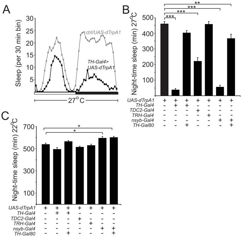
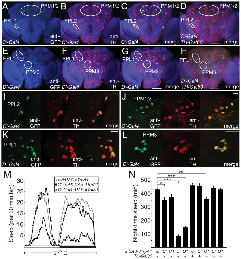
Background: The neuronal circuitry underlying sleep is poorly understood. Although dopamine (DA) is thought to play a key role in sleep/wake regulation, the identities of the individual DA neurons and their downstream targets required for this process are unknown.

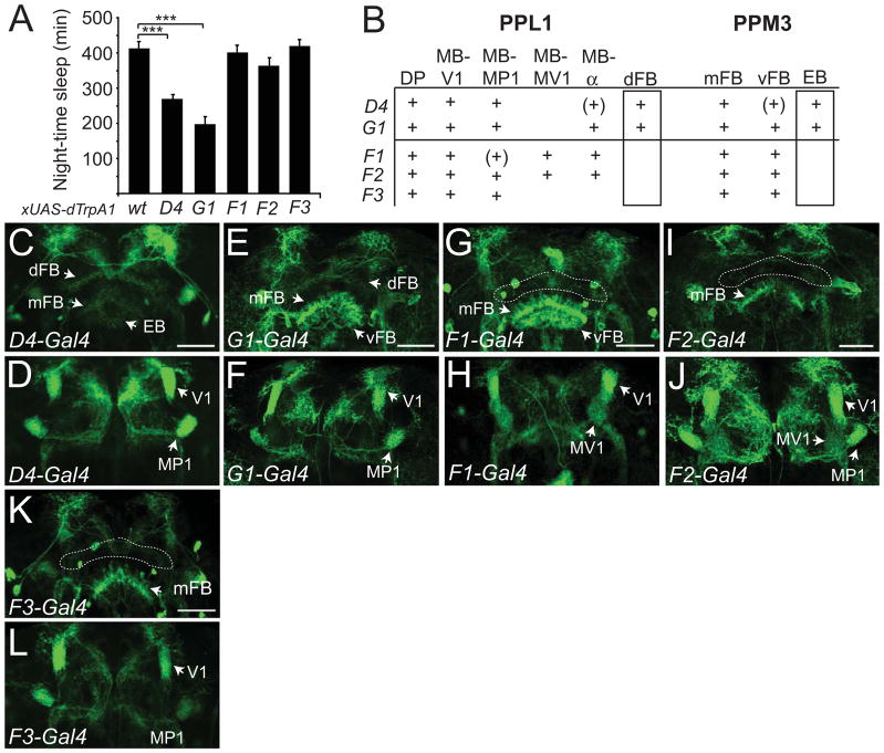
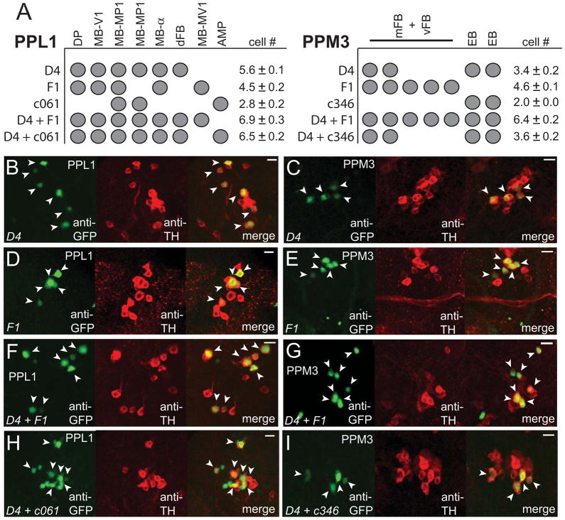
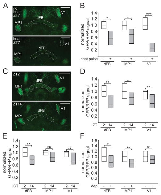
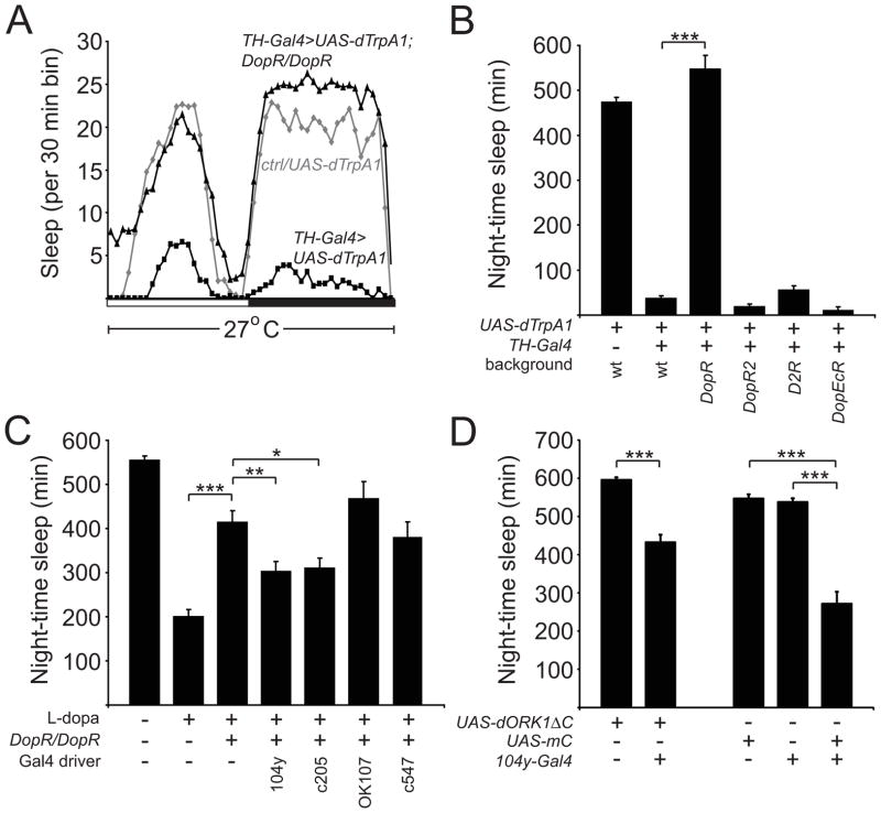
Results: Here, we identify a DA neuron in each PPL1 cluster that promotes wakefulness in Drosophila. Imaging data suggest that the activity of these neurons is increased during wakefulness, consistent with a role in promoting arousal. Strikingly, these neurons project to the dorsal fan-shaped body, which has previously been shown to promote sleep. The reduced sleep caused by activation of DA neurons can be blocked by loss of DopR, and restoration of DopR expression in the fan-shaped body can rescue the wake-promoting effects of DA in a DopR mutant background.

Conclusions: These experiments define a novel arousal circuit at the single-cell level. Because the dorsal fan-shaped body promotes sleep, these data provide a key link between wake and sleep circuits. Furthermore, these findings suggest that inhibition of sleep centers via monoaminergic signaling is an evolutionarily conserved mechanism to promote arousal.


Figures

Figure 1
Figure 1. Dopamine Signaling is a Key Arousal Pathway in Drosophila
(A) Sleep profile of UAS-dTrpA1/+ (gray diamonds) vs TH-Gal4/UAS-dTrpA1 (black squares) flies at 27°C in 12:12 LD, plotted in 30 min bins. White and black bars denote light and dark periods, respectively. Night-time (B) sleep amount at 27°C and (C) sleep amount at 22°C plotted for UAS-dTrpA1/+ (n=54), TH-Gal4/UAS-dTrpA1 (n=36), TH-Gal80/+; TH-Gal4/UAS-dTrpA1 (n=66), TDC2-Gal4/+; UAS-dTrpA1 (n=37), TRH-Gal4/UAS-dTrpA1 (n=42), nsyb-Gal4/+; UAS-dTrpA1/+ (n=50), and nsyb-Gal4/TH-Gal80; UAS-dTrpA1/+ (n=34) flies. In this and subsequent figures, error bars represent SEM. “*”, “**”, and “***” denote P<0.05, P<0.01, and P<0.001 respectively.
Figure 2
Figure 2. Analysis of Novel DA Gal4 Drivers Suggests that the PPL1 and/or PPM3 Clusters Mediate the Arousing Effects of DA
Maximum projection confocal images of the posterior half of whole mount adult brains of flies expressing GFP-nls driven by TH-C-Gal4 (A–D) or TH-D-Gal4 (E–H) in the absence (A–C, E–G) or presence (D, H) of TH-Gal80. Single higher magnification confocal sections showing PPL2 (I) and PPM1/2 (J) in TH-C-Gal4 flies expressing GFP-nls, and PPL1 (K) and PPM3 (L) in TH-D-Gal4 flies expressing GFP-nls. Brains are immunostained with nc82, anti-GFP, and anti-TH antibodies. Scale bars denote 100 μm (A–H) and 20 μm (I–L). (M) Sleep profile of UAS-dTrpA1/+ (gray diamonds), TH-C-Gal4/+; UAS-dTrpA1/+ (black squares), and TH-D-Gal4/+; UAS-dTrpA1/+ (black triangles) flies at 27°C in 12:12 LD, plotted in 30 min bins. White and black bars denote light and dark periods, respectively. (N) Night-time sleep plotted for UAS-dTrpA1/+ (n=63), TH-C-Gal4/+; UAS-dTrpA1/+ (n=27), TH-C1-Gal4/+; UAS-dTrpA1/+ (n=38), TH-D-Gal4/+; UAS-dTrpA1/+ (n=40), TH-D1-Gal4/+; UAS-dTrpA1/+ (n=46), TH-Gal80/+; UAS-dTrpA1/+ (n=44), TH-C-Gal4/TH-Gal80; UAS-dTrpA1/+ (n=32), TH-C1-Gal4/TH-Gal80; UAS-dTrpA1/+ (n=49), TH-D-Gal4/TH-Gal80; UAS-dTrpA1/+ (n=41), TH-D1-Gal4/TH-Gal80; UAS-dTrpA1/+ (n=51) flies. “wt” denotes background control.
Figure 3
Figure 3. Activation of Gal4 Drivers with Restricted Expression in PPL1 and/or PPM3 Suggests that DA cells Projecting to the Dorsal Fan-shaped Body or Ellipsoid Body Promote Wakefulness
(A) Night-time sleep for UAS-dTrpA1/+ (n=56), TH-D4-Gal4/UAS-dTrpA1 (n=71), TH-G1-Gal4/UAS-dTrpA1 (n=40), TH-F1-Gal4/UAS-dTrpA1 (n=62), TH-F2-Gal4/UAS-dTrpA1 (n=51), and TH-F3-Gal4/UAS-dTrpA1 (n=42) flies at 27°C. (B) Identities of the projections from the PPL1 and PPM3 clusters for the TH-D4, TH-G1, TH-F1, TH-F2, TH-F3 lines, with naming as described in Table S1. “+” denotes presence of projection, and “(+)” denotes faint staining for projection. Confocal images of brains immunostained with anti-GFP of TH-D4 (C, D), TH-G1 (E, F), TH-F1 (G, H), TH-F2 (I, J), or TH-F3 (K, L) driving expression of UAS-mCD8-GFP; UAS-mCD8-GFP. Images for (C, E, G, I, K) and (D, F, H, J, L) consist of maximum projections of a ~4 μm section through the middle or anterior third of the adult brain to highlight the central complex and mushroom bodies, respectively. “dFB”, “EB”, “mFB”, “vFB”, “V1”, “MP1”, “MV1” indicate projections for the dorsal fan-shaped body, ellipsoid body, middle fan-shaped body, ventral fan-shaped body, and the V1, MP1, or MV1 regions of the mushroom bodies. Dashed lines outline the location for the dFB in (G), (I), and (K). Scale bar denotes 50μm.
Figure 4
Figure 4. A Single Neuron from the PPL1 Subgroup, which Projects to the Dorsal Fan-shaped Body, Promotes Wakefulness
(A) Identities of the TH+ cells in the PPL1 and PPM3 clusters in the indicated Gal4 driver lines. Mean cell counts for PPL1 and PPM3 are shown for TH-D4-Gal4 (n=13 hemispheres), TH-F1-Gal4 (n=14), c061-Gal4 (n=5), c346-Gal4 (n=3), TH-D4-Gal4/TH-F1-Gal4 (n=11), c061-Gal4; TH-D4-Gal4 (n=8), and c346-Gal4; TH-D4-Gal4 (n=9) flies expressing GFP-nls. (B–I) Single confocal sections showing the TH+ cells in the PPL1 or PPM3 clusters for the indicated driver lines expressing GFP-nls. Arrowheads indicate GFP+ TH+ cells. Scale bar denotes 10 μm.
Figure 5
Figure 5. Imaging experiments using ANF-GFP suggest the PPL1-dFB neurons are more active during the day and during wakefulness
(A) Whole-mount brains from flies expressing ANF-GFP, myr-RFP, and dTrpA1, under control of TH-D4-Gal4. Images consist of maximum projections of a 12 μm section through the middle of the brain to highlight the mushroom body and central complex projections, with either no heat pulse (“no heat”) or 5 hr 32°C heat pulse (“heat”) from ZT2–ZT7. “dFB”, “MP1”, and “V1” indicate projections to the dorsal fan-shaped body or MP1 and V1 regions of the mushroom bodies, respectively. Dashed lines highlight these projections. (B) GFP/RFP signal for these flies is represented as simplified box plots, where the line inside the box indicates the median, and the top and bottom represent 75th and 25th percentiles, respectively. n=7 for no heat pulse and n=12 for heat pulse conditions. (C) Whole-mount brains from flies expressing ANF-GFP and myr-RFP under control of TH-D4-Gal4 at ZT2 or ZT14. GFP/RFP signal is shown as simplified box plots at ZT2 (n=20) and ZT14 (n=21) (D), CT2 (n=15) and CT14 (n=20) (E), or no sleep deprivation (no dep, n=22) and sleep deprivation from ZT12–24 (dep, n=31) (F) conditions. Scale bar denotes 50 μm.
Figure 6
Figure 6. DopR Mediates the Arousing Effects of DA and is Required in the Fan-shaped Body to Promote Wakefulness
(A) Sleep profile of UAS-dTrpA1/+ (gray diamonds), TH-Gal4/UAS-dTrpA1 (black squares), and TH-Gal4, DopR/UAS-dTrpA1, DopR flies (black triangles) at 27°C in 12:12 LD, plotted in 30 min bins. White and black bars denote light and dark periods, respectively. (B) Night-time sleep for UAS-dTrpA1/+ (n=62), TH-Gal4/UAS-dTrpA1 (n=81), TH-Gal4, DopR/UAS-dTrpA1, DopR (n=31), TH-Gal4, DopR2/UAS-dTrpA1, DopR2 (n=31), D2R;;TH-Gal4/UAS-dTrpA1 (n=41), and TH-Gal4, DopEcR/UAS-dTrpA1, DopEcR (n=32) flies at 27°C. (C) Night-time sleep for control flies not fed L-dopa (n=64) and control (n=145), DopR/DopR (n=69), 104y-Gal4/+; DopR/DopR (n=82), c205-Gal4, DopR/DopR or c205-Gal4, DopR (n=67), DopR/DopR; OK107/+ (n=26), and c547-Gal4, DopR/DopR (n=32) flies fed 4 mg/ml L-dopa at 25°C. (D) Night-time sleep for UAS-dORK1ΔC/+ (n=59), 104y-Gal4/+; UAS-dORK1ΔC/+ (n=35), UAS-mC*/+ (n=62), 104y-Gal4/+ (n=31), and 104y-Gal4/UAS-mC* (n=37) flies at 25°C.

References

    1. Saper CB, Fuller PM, Pedersen NP, Lu J, Scammell TE. Sleep state switching. Neuron. 2010;68:1023–1042. - PMC - PubMed
    1. Tanaka C, Ishikawa M, Shimada S. Histochemical mapping of catecholaminergic neurons and their ascending fiber pathways in the rhesus monkey brain. Brain Res Bull. 1982;9:255–270. - PubMed
    1. Hendricks JC, Finn SM, Panckeri KA, Chavkin J, Williams JA, Sehgal A, Pack AI. Rest in Drosophila is a sleep-like state. Neuron. 2000;25:129–138. - PubMed
    1. Shaw PJ, Cirelli C, Greenspan RJ, Tononi G. Correlates of sleep and waking in Drosophila melanogaster. Science. 2000;287:1834–1837. - PubMed
    1. Cirelli C, Bushey D, Hill S, Huber R, Kreber R, Ganetzky B, Tononi G. Reduced sleep in Drosophila Shaker mutants. Nature. 2005;434:1087–1092. - PubMed

Publication types

MeSH terms

Substances