. 2013 Jan;25(1):178-89.
doi: 10.1016/j.cellsig.2012.09.024. Epub 2012 Sep 27.

Cripto-1 enhances the canonical Wnt/β-catenin signaling pathway by binding to LRP5 and LRP6 co-receptors

Affiliations

Cripto-1 enhances the canonical Wnt/β-catenin signaling pathway by binding to LRP5 and LRP6 co-receptors

Tadahiro Nagaoka et al. Cell Signal. 2013 Jan.

Abstract

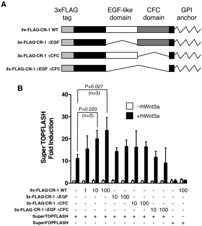
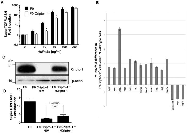
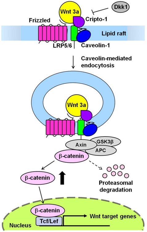
Cripto-1 is implicated in multiple cellular events, including cell proliferation, motility and angiogenesis, through the activation of an intricate network of signaling pathways. A crosstalk between Cripto-1 and the canonical Wnt/β-catenin signaling pathway has been previously described. In fact, Cripto-1 is a downstream target gene of the canonical Wnt/β-catenin signaling pathway in the embryo and in colon cancer cells and T-cell factor (Tcf)/lymphoid enhancer factor binding sites have been identified in the promoter and the first intronic region of the mouse and human Cripto-1 genes. We now demonstrate that Cripto-1 modulates signaling through the canonical Wnt/β-catenin/Tcf pathway by binding to the Wnt co-receptors low-density lipoprotein receptor-related protein (LRP) 5 and LRP6, which facilitates Wnt3a binding to LRP5 and LRP6. Cripto-1 functionally enhances Wnt3a signaling through cytoplasmic stabilization of β-catenin and elevated β-catenin/Tcf transcriptional activation. Conversely, Wnt3a further increases Cripto-1 stimulation of migration, invasion and colony formation in soft agar of HC11 mouse mammary epithelial cells, indicating that Cripto-1 and the canonical Wnt/β-catenin signaling co-operate in regulating motility and in vitro transformation of mammary epithelial cells.


Figures

Fig. 1
Fig. 1
Cripto-1 enhances Wnt3a induced SuperTOPFLASH luciferase activity in 293T cells. (A) Full-length (CR-1 WT) and deletion constructs (CR-1 ΔEGF lacking the EGF-like domain, CR-1 ΔCFC lacking the CFC domain, CR-1 ΔEGF ΔCFC lacking both EGF and CFC domains) of human Cripto-1 in a 3×-FLAG expression vector. (B) SuperTOPFLASH luciferase reporter assay in 293T cells transiently transfected with different concentrations of full-length 3×-FLAG CR-1 WT plasmid (1, 10 and 100 ng) or with 3×-FLAG CR-1 ΔEGF, 3×-FLAG CR-1 ΔCFC, 3×-FLAG CR-1 ΔEGF ΔCFC deletion constructs (10 and 100 ng) together with 50 ng of SuperTOPFLASH luciferase vector. Cells were subsequently treated with rhWnt3a protein (50 ng/ml) for 16-20 hr. Cells were also transfected with 50 ng of SuperFOPFLASH luciferase vector and subsequently stimulated with rhWnt3a. These results are the mean ±SD of triplicates from one of three separate experiments.
Fig. 2
Fig. 2
Cripto-1 lacking GPI-anchor does not enhance the canonical Wnt/β-catenin signaling pathway stimulated by Wnt3a in 293T cells. (A) Full-length (CR-1 WT) and GPI-anchor signal sequence deletion mutant (CR-1 ΔC) of human Cripto-1 in a 3×FLAG expression vector. (B) SuperTOPFLASH luciferase assay in 293T cells transiently transfected with different concentrations of 3×FLAG-CR-1 WT or 3×FLAG-CR-1 ΔC (10 and 100 ng) together with 50 ng of SuperTOPFLASH luciferase vector. Cells were subsequently treated with 50 ng/ml of rhWnt3a protein for 16-20 hr. These results are the mean ±SD of triplicates from one of three separate experiments.
Fig. 3
Fig. 3
Interaction between CR-1 and Wnt ligand or Wnt receptors. Mouse Wnt3a-HA, human Fz8 CRD-Fc, human LRP5ΔC-myc and full-length 3×FLAG-CR-1 WT expression plasmids were co-transfected into 293T cells. After 24 h, cells were harvested and lysed in RIPA buffer. The cell lysates were treated with 10 μl of anti-FLAG M2 affinity gel for 2 h at 4 °C. The Affinity gels were washed with RIPA buffer for 4 times. Proteins were then eluted by 2× Laemmli SDS sample buffer and separated by SDS-PAGE. Western blotting was carried out using following antibodies: anti-FLAG, anti-myc, anti-HA and anti-human IgG-HRP.
Fig. 4
Fig. 4
Cripto-1 binds to LRP5 and LRP6 in 293T cells. 293T cells were co-transfected with LRP-5-tGFP or LRP6-GFP expression vectors together with Cripto-1 full-length or deleted expression plasmids. 3×-FLAG Cripto-1 was immunoprecipated using an anti-FLAG mouse monoclonal antibody and the immunoprecipitated proteins were analyzed by Western blot with anti-tGFP, anti-GFP and anti-FLAG mouse monoclonal antibodies (A and C). LRP5-tGFP or LRP6-GFP were immunoprecipitated with anti-tGFP or anti-GFP mouse monoclonal antibody and immunoprecipitated proteins were analyzed by Western blot with anti-tGFP, anti-GFP and anti-FLAG mouse monoclonal antibodies (B and D). Cell lysates of 293T cells transiently transfected with various expression vectors as described above were analyzed by Western blot for Cripto-1 using an anti-FLAG antibody, for LRP5 using anti-tGFP antibody and for LRP6 using anti-GFP antibody (A through D).
Fig. 5
Fig. 5
Cripto-1 binds to LRP5 and LRP6 co-receptors on the cell surface. 293T cells transiently transfected with Cripto-1, LRP5 and/or LRP6 expression plasmids were incubated with 2mM NHS-PEG4-Biotin and then lysed using RIPA buffer. Following immunoprecipitation with anti-FLAG or anti-GFP monoclonal antibodies, cell surface biotinylated proteins were isolated by adding streptavidin agarose beads to the immunoprecipitated proteins for 3 h at 4 °C. A and C show co-immunoprecipitation of LRP5-tGFP (A) or LRP6-GFP (C) with 3×-FLAG CR-1 WT only in the presence of biotin. B and D show co-immunoprecipitation of 3×-FLAG CR-1 WT with LRP5-tGFP (B) or LRP6-GFP (D) only in the presence of biotin. Cell lysates of immunoprecipitated samples were also analyzed by Western blot to ensure expression of transfected plasmids. (E) Endogenous Cripto-1 and LRP6 co-immunoprecipitate in NCCIT human embryonal carcinoma cells. NCCIT protein lysates were immunoprecipitated with an anti-Cripto-1 (V-17) rabbit polyclonal antibody and probed with an LRP6 monoclonal antibody (E) or immunoprecipitated with an anti-LRP6 monoclonal antibody and probed with and anti-Cripto-1 rabbit polyclonal antibody (F). * indicates the immunoglobulin light chain.
Fig. 6
Fig. 6
Cripto-1 and LRP6 co-localize on the cell membrane. 293T/Cripto-1 cells transiently transfected with LRP6-GFP expression vector were stained with anti-LRP6 (green) and anti-Cripto-1 (red) antibodies. Nuclei were visualized by Hoechst 33258 (blue). The merged image shows significant co-localization of Cripto-1 and LRP6 on the cell membrane. The arrows indicate clear examples of co-localization.
Fig. 7
Fig. 7
β-catenin stabilization and LRP5/6 phosphorylation in F9 and F9 Cripto-1-/- cells treated with rhWnt3a. (A) F9 and F9 Cripto-1-/- cells were stimulated with different concentrations of rhWnt3a and activated β-catenin was immunoprecipitated with GST-E-cadherin. Glutathione agarose beads were then added to immunoprecipitated proteins and eluted proteins were analyzed by Western blot using an anti-β-catenin antibody. Densitometric analysis of immunoprecipitated β-catenin was performed with Image J program. Protein lysates were analyzed by Western blot for β-catenin, Cripto-1 and β-actin expression (B) Western blot analysis of phosphorylated LRP5/6 in F9 and F9 Cripto-1-/- cells after rhWnt3a treatment. Density ratio (P/T) corresponds to phosphorylated LRP6 (P-LRP6) divided by total LRP6 (LRP6).
Fig. 8
Fig. 8
Wnt/ β-catenin signaling in F9 and F9 Cripto-1-/- cells. (A) Luciferase reporter assay in F9 and F9 Cripto-1-/- cells transiently transfected with Tcf/Lef responsive SuperTOPFLASH luciferase reporter vector. After transfection, cells were stimulated with different concentrations of rhWnt3a (B) Wnt signaling molecules real time PCR array in F9 and F9 Cripto-1-/- cells. Cells were treated with/without rhWnt3a (100 ng/ml) for 18 h, cDNA was synthesized from total RNA and real time PCR array was performed according to manufacturer's instructions. (C) and (D) Re-expression of Cripto-1 in F9 Cripto-1-/- cells partially rescues sensitivity of F9 Cripto-1-/- cells to rhWnt3a stimulation. (C) A lentiviral Cripto-1 expression vector or a control empty vector were infected into F9 Cripto-1-/- cells, stably expressing lentiviral vectors cells were established and Cripto-1 expression was assessed by Western blot analysis. (D) F9 Cripto-1-/- cells, stably expressing a lentiviral Cripto-1 expression vector or a control empty vector, were compared to F9 control cells in a SuperTOPFLASH luciferase assay after rhWnt3a stimulation (10 ng/ml). P value was calculated using Student's t test.
Fig. 9
Fig. 9
Activation of the canonical Wnt/β-catenin signaling pathway is independent of a Smad2/3 signaling pathway in F9 cells. (A) Western blot analysis for phospho Smad2 (P-Smad2) and total Smad2 in F9 and F9 Cripto-1-/- cells stimulated with rhWnt3a (10 ng/ml) in the presence or absence of SB431542 inhibitor. Density ratio indicates densitometric values of phosphorylated Smad2/total Smad2. (B) SuperTOPFLASH luciferase reporter assay and (C) (n2)7-luc reporter assay in F9 and F9 Cripto-1-/- cells. Cells were incubated in the presence of the Alk4/5/7 inhibitor SB431542 to block activation of the Smad2/Smad3 signaling pathway.
Fig. 10
Fig. 10
F9 cells are more resistant to Dkk1 inhibition of the Wnt/β-catenin signaling pathway than F9 Cripto-1-/- cells. (A) SuperTOPFLASH assay in F9 and F9 Cripto-1-/- cells treated with rhWnt3a (100 ng/ml) alone or in combination with different concentration of rmDkk1. (B) Dkk1 mRNA expression in F9 and F9 Cripto-1-/- cells as assessed by RT-PCR.
Fig. 11
Fig. 11
Wnt3a enhances Cripto-1 stimulated migration, invasion and growth in soft agar of HC11 mouse mammary epithelial cells. (A) Western blot analysis for Cripto-1 and β-actin in HC11 control cells (WT) and HC11 stably expressing Cripto-1 (HC11/Cripto-1). Migration (B) and invasion (C) assays of HC11 and HC11/Cripto-1 in the presence or absence of rhWnt3a protein (50 ng/ml). (D) Growth in soft agar of HC11 and HC11/Cripto-1 cells in the presence or absence of rhWnt3a (50 ng/ml). Colonies were counted after 2 weeks. * P <0.05 as compared to HC11 untreated cells; **P< 0.05 as compared to HC11/Cripto-1 untreated cells.
Fig. 12
Fig. 12
Proposed model of Cripto-1 modulation of the Wnt/β-catenin signaling pathway. Cripto-1 can regulate the Wnt/β-catenin signaling pathway through the binding of LRP5/6, which also bind to caveolin-1 in the lipid raft as well as Cripto-1. Cripto-1 facilitates the phosphorylation of LRP5/6 induced by Wnt3a. The phosphorylated LRP6 leads to recruitment of the Axin complex (Axin, APC and GSK3β) to the Wnt receptor complex. The receptor complex is then internalized via a caveolin-mediated route to induce the cytoplasmic accumulation of β-catenin. β-catenin can then form a complex with Tcf/Lef and activate the transcription of target genes after translocation to the nucleus. Cripto-1 also might interfere with the internalization of LRP5/6 that is facilitated by Dkk1, which is a potent inhibitor of the Wnt/β-catenin signaling pathway.

References

    1. Dono R, Montuori N, Rocchi M, De Ponti-Zilli L, Ciccodicola A, Persico MG. Am J Hum Genet. 1991;49(3):555–565. - PMC - PubMed
    1. Ciccodicola A, Dono R, Obici S, Simeone A, Zollo M, Persico MG. Embo J. 1989;8(7):1987–1991. - PMC - PubMed
    1. Minchiotti G, Parisi S, Liguori G, Signore M, Lania G, Adamson ED, Lago CT, Persico MG. Mech Dev. 2000;90(2):133–142. - PubMed
    1. de Castro NP, Rangel MC, Nagaoka T, Salomon DS, Bianco C. Future Oncol. 2010;6(7):1127–1142. - PubMed
    1. Bianco C, Rangel MC, Castro NP, Nagaoka T, Rollman K, Gonzales M, Salomon DS. Am J Pathol. 2010;177(2):532–540. - PMC - PubMed

Publication types

MeSH terms