. 2012 Nov;44(11):1265-71.
doi: 10.1038/ng.2426. Epub 2012 Sep 30.

Alterations of the CIB2 calcium- and integrin-binding protein cause Usher syndrome type 1J and nonsyndromic deafness DFNB48

Affiliations

Alterations of the CIB2 calcium- and integrin-binding protein cause Usher syndrome type 1J and nonsyndromic deafness DFNB48

Saima Riazuddin et al. Nat Genet. 2012 Nov.

Erratum in

Abstract

Sensorineural hearing loss is genetically heterogeneous. Here, we report that mutations in CIB2, which encodes a calcium- and integrin-binding protein, are associated with nonsyndromic deafness (DFNB48) and Usher syndrome type 1J (USH1J). One mutation in CIB2 is a prevalent cause of deafness DFNB48 in Pakistan; other CIB2 mutations contribute to deafness elsewhere in the world. In mice, CIB2 is localized to the mechanosensory stereocilia of inner ear hair cells and to retinal photoreceptor and pigmented epithelium cells. Consistent with molecular modeling predictions of calcium binding, CIB2 significantly decreased the ATP-induced calcium responses in heterologous cells, whereas mutations in deafness DFNB48 altered CIB2 effects on calcium responses. Furthermore, in zebrafish and Drosophila melanogaster, CIB2 is essential for the function and proper development of hair cells and retinal photoreceptor cells. We also show that CIB2 is a new member of the vertebrate Usher interactome.


Figures

Figure 1
Figure 1
Pedigrees of USH1J/DFNB48 families. One USH1J and four of 57 NSHI DFNB48 families segregating CIB2 (NM_006383) mutant alleles. Filled symbols represent affected individuals and a double horizontal line is a consanguineous marriage. Haplotypes for selected individuals of families PKDF356 and PKDF282 indicate the smallest linkage interval. The proximal breakpoint (arrow) is defined by affected individual VI:3 of family PKDF356 at marker D15SA985 (78.13 Mb). The distal breakpoint (arrowhead) is defined by unaffected individual V:5 (PKDF282) at D15SA975 (78.56 Mb). CIB2 mutant alleles [c.272T>C (p.Phe91Ser), c.297C>G (p.Cys99Trp) and c.297C>G (p.Ile123Thr)] co-segregate with NSHI phenotype in PKDF356, PKDF282, DEM4225 and family-802, respectively. The USH1 phenotype of family PKDF117 co-segregates with c.192G>C (p.Glu64Asp) mutation of CIB2. These four recessive mutations co-segregate with deafness or deaf-blindness while carriers have normal hearing.
Figure 2
Figure 2
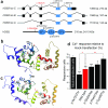
CIB2 isoforms, molecular models, and functional effects of mutations. (a) Human CIB2 has six exons encoding three isoforms. Non-coding, EF-hand domains and other coding regions of exons are denoted by grey, blue and black boxes, respectively. (b) Molecular models using template 1XO5.PDB crystal structure of Ca2+-CIB1. (c) Model of CIB2 using a template for NMR structure of CIB1 bound to αIIβ integrin peptide. (b-c) The backbone ribbon is color-coded blue (N-terminus) to red (C-terminus) and two Ca2+-ions are blue spheres. (d) Ca2+ responses in COS-7 cells transfected with five different DsRed-tagged CIB2 constructs. Data normalized to average response of mock-transfections; shown as mean ± SE. Asterisks indicate statistical significance: ***, p<0.001; *, p<0.05. None of the four missense mutations resulted in noticeable changes of CIB2 distribution (not shown). p.Cys99Trp abolished CIB2’s ability to decrease sensitivity of antagonist-induced Ca2+ release from the cell, while p.Ile123Thr enhanced this ability.
Figure 3
Figure 3
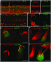
CIB2 localization in hair cells of the organ of Corti and vestibular sensory epithelia. (ac) Localization of CIB2 (green) in the inner hair cell stereocilia. F-actin is visualized by rhodamine-phalloidin (red). (b, c) shows boxed area in a at a higher magnification, separated in red (b, rhodamine-phalloidin) and green (c, CIB2) channels. The region of interest in b and c covered the tips of the second row stereocilia and was used to measure integrated intensity of fluorescent signal (Supplementary Table 6). (d) CIB2 (green) is present in stereocilia of outer hair cells. (e, f). CIB2 (green) in vestibular hair cell stereocilia cluster in patches around the actin core (red). Most of the CIB2 patches (f, green channel alone) are observed near or at the tips of stereocilia. (g, i) Gene gun transfection of the organ of Corti hair cells with CIB2-GFP shows predominant targeting of CIB2 to stereocilia tips, particularly to the tips of the shorter row of inner (g) and outer (i) hair cell stereocilia. (h, j) Vestibular hair cell transfected with CIB2-GFP show greater concentration of CIB2-GFP (green) to the tips of shorter row stereocilia (red). Scale bars in all of the panels are 5 μm, except d. Scale bar in d is 10 μm.
Figure 4
Figure 4
CIB2 homodimerizes and also interacts with whirlin and myosin VIIa (a) Possible CIB2 interactome. Myosin VIIa might be interacting directly or through an intermediate protein. (b) CIB2 homodimerizes. Lysates from HEK293 cells transfected with CIB2-GFP and tdTomato-CIB2 expression constructs were co-immunoprecipitated with anti-GFP antibody. Precipitates were immuno-blotted with CIB2 and GFP antibody. (c) CIB2 and myosin VIIa interact. Lysates from HEK293 cells transfected with GFP-MyoVIIa and tdTomato-CIB2 constructs were co-immuno-precipitated with anti-GFP antibody. Precipitates were immuno-blotted with CIB2 and GFP antibodies. (d) CIB2 and whirlin interact. Lysates from HEK293 cells co-transfected with CIB2-GFP and DsRed-whirlin were co-immunoprecipitated with an anti-GFP antibody. Precipitates were immuno-blotted with CIB2 and whirlin antibodies. As negative control, we transfected CIB2-GFP either DsRed or tdTomato empty vectors and did not observe an interaction (Supplementary Fig. 10b-c). In this assay CIB2 does not interact with harmonin, cadherin 23, protocadherin15-CD1, -CD2 and –CD3, Sans, usherin, vlgr1 or clarin-1.
Figure 5
Figure 5
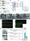
Suppression of cib2 expression produces developmental defects in zebrafish embryos. (a - b) Embryos injected with morpholino (MO) against cib2 have developmental defects including micro-ophthalmia, curled tail, hypo-pigmentation and an edematous heart. (c) Acoustic startle reflex of 5-day-old morphants. A significant percentage of cib2 MO injected larvae either did not respond to acoustic stimuli, indicating HI, or were unable to remain upright while swimming, indicative of a balance defect, shown as mean ± SE (d) Scanning electronic microscopic (SEM) imaging revealed normal morphology of hair bundle in the neuromast. (e) Shows boxed area in d at a higher magnification. (f - i) SEM image revealed normal looking hair bundles in the neuromast cells in class I and class II morphants. (g, i) High-resolution imaging of neuromast present in the class I and class II morphants (boxed areas in f and h) showed the intact hair bundle links connecting the different rows. (j, k) Complete absence of neuromasts at the lateral lines of class III and IV cib2 morphants. Scale bars: 5 μM for panel j, 3 μM for d and f, 2 μM for h and k, 500 nM for e, g and i. (l – o) Inhibition of microphonic potentials in zebrafish lateral line by cib2 morpholino. (l) Comparison of FM1–43 labeling in larvae from all four classes at 72 hpf showed a marked reduction of uptake in cib2 morphants. Arrow and arrowheads indicate the neuromast cells. (m), Bright field image of a neuromast in a zebrafish injected with control morpholino and with cib2 morpholino. (n) Microphonic potentials in neuromasts of non-injected wild type fish (top trace), fish injected with control morpholino (second trace), and fish injected with cib2 morpholino (third trace). The bottom trace indicates 2 μm peak-to-peak (p-p) stimulation. (o) Average peak-to-peak amplitude of microphonic potential in wild type, control, and cib2 morphants. Number of neuromasts: 17, wild type; 11, control; 20, CIB2. Data are shown as Mean ± SE. Asterisks indicate statistical significance: p<0.001.
Figure 6
Figure 6
Physiological and morphological changes in Drosophila dCib2 deficient retinas. (a) Electroretinogram illustrating the photo response to a 5 second white light pulse. dCib2RNAi flies show a reduction in response amplitude of over 30% compared to control flies. (b) The amplitude reduction is significant (student t-test), with a p-value of 0.02 for 5 second pulses, and p=0.00005 for 300ms pulses. (c) An example of a 40Hz series (train) of light pulses illustrates that dCib2RNAi flies are unable to follow fast trains of light pulses as well as control flies. At this frequency, the electric signal of control flies closely follows all light pulses, while dCib2RNAi flies skip the majority of pulses and show only a very weak response to a few pulses. (d) Response amplitudes in dCib2RNAi flies decreased to the noise levels at lower frequencies than do the responses in control flies. (e) In contrast to control flies, dCib2RNAi flies are unable to maintain a persistent response during prolonged stimulation at a low frequency (1.7Hz). (f) On average, the response strength of dCIB2RNAi flies becomes indistinguishable from noise levels after the first 22 pulses of stimulation. (g) Analysis of dCib2RNAi retinas by water immersion (top two panel sets) or thin plastic sections (bottom panel set) reveal little to no differences from controls when raised in 12hr:12hr light:dark cycles (not shown) or in complete darkness. However, significant degeneration is observed when flies are raised in constant light for 5 days, indicating that Cib2 is necessary to prevent light-induced retinal degeneration.

Comment in

References

    1. Ahmad J, et al. DFNB48, a new nonsyndromic recessive deafness locus, maps to chromosome 15q23-q25.1. Hum Genet. 2005;116:407–12. - PubMed
    1. Ahmed ZM, Riazuddin S, Khan SN, Friedman PL, Friedman TB. USH1H, a novel locus for type I Usher syndrome, maps to chromosome 15q22-23. Clin Genet. 2009;75:86–91. - PMC - PubMed
    1. Adzhubei IA, et al. A method and server for predicting damaging missense mutations. Nature methods. 2010;7:248–9. - PMC - PubMed
    1. Schwarz JM, Rodelsperger C, Schuelke M, Seelow D. MutationTaster evaluates disease-causing potential of sequence alterations. Nature methods. 2010;7:575–6. - PubMed
    1. Gentry HR, et al. Structural and biochemical characterization of CIB1 delineates a new family of EF-hand-containing proteins. J Biol Chem. 2005;280:8407–15. - PubMed

Publication types

MeSH terms

Substances

Supplementary concepts