. 2012 Nov;122(11):3914-8.
doi: 10.1172/JCI63065.

Intrahepatic cholangiocarcinoma can arise from Notch-mediated conversion of hepatocytes

Affiliations

Intrahepatic cholangiocarcinoma can arise from Notch-mediated conversion of hepatocytes

Sayaka Sekiya et al. J Clin Invest. 2012 Nov.

Abstract

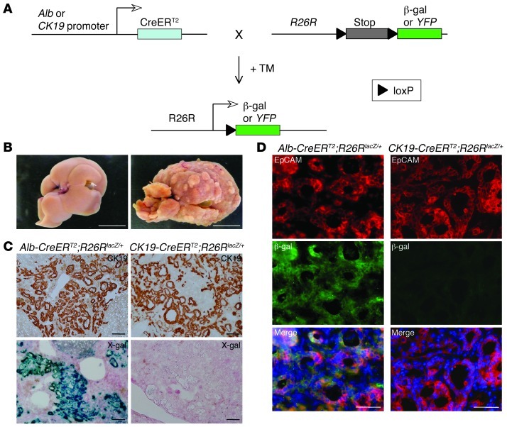
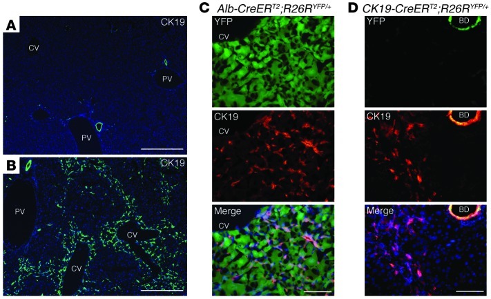
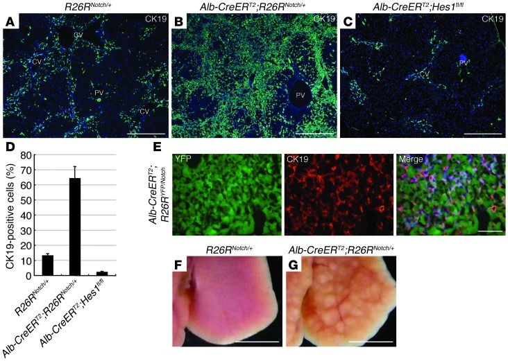
Intrahepatic cholangiocarcinoma (ICC) is the second most common primary malignancy in the liver. ICC has been classified as a malignant tumor arising from cholangiocytes; however, the co-occurrence of ICC and viral hepatitis suggests that ICC originates in hepatocytes. In order to determine the cellular origin of ICC, we used a mouse model of ICC in which hepatocytes and cholangiocytes were labeled with heritable, cell type–specific reporters. Our studies reveal that ICC is generated by biliary lineage cells derived from hepatocytes, rather than cholangiocytes. Additionally, we found that Notch activation is critical for hepatocyte conversion into biliary lineage cells during the onset of ICC and its subsequent malignancy and progression. These findings will help to elucidate the pathogenic mechanism of ICC and to develop therapeutic strategies for this refractory disease.


Figures

Figure 1
Figure 1. Primitive ductules formed in ICC are derived from hepatocytes, rather than cholangiocytes.
(A) Experimental procedure to follow the lineage of hepatocytes and cholangiocytes in mouse liver. In the presence of TM, CreERT2 expressed from the Alb or CK19 genomic locus translocates into the nucleus and removes the loxP-flanked stop cassette from the R26R allele, leading to permanent heritable expression of the β-gal or YFP gene. (B) Representative images of liver from mice that drank normal water (left) or TAA-containing water (right) for 30 weeks. Tumoral macronodules only formed after TAA administration. (C and D) Immunohistochemical staining of CK19 (C, top row), X-gal staining (C, bottom row), and co-immunofluorescence staining of β-gal and EpCAM (D) were conducted for neoplastic nodules formed in the liver of Alb-CreERT2;R26RlacZ/+ mice and CK19-CreERT2;R26RlacZ/+ mice after 30 weeks’ TAA administration. DNA was stained with DAPI. Scale bars: 10 mm (B), 100 μm (C), and 50 μm (D).
Figure 2
Figure 2. Hepatocytes on the periphery of central veins in the hepatic lobules are converted into biliary lineage cells at the onset of ICC.
(AD) Immunofluorescence staining of CK19 was conducted in the liver of control wild-type mice (A) and the liver of wild-type mice (B), Alb-CreERT2;R26RYFP/+ mice (C), and CK19-CreERT2;R26RYFP/+ mice (D) after 14 weeks of TAA administration. CV, cetral vein; PV, portal vein; BD, bile duct. DNA was stained with DAPI. Scale bars: 500 μm (A and B) and 100 μm (C and D).
Figure 3
Figure 3. Notch signal activation is significant not only for conversion of hepatocytes into biliary lineage cells at the onset of ICC, but also the malignancy and progression of ICC.
(AC) Immunofluorescence staining of CK19 was conducted in the liver of R26RNotch/+ mice (A), Alb-CreERT2;R26RNotch/+ mice (B), and Alb-CreERT2;Hes1fl/fl mice (C) after 14 weeks of TAA administration. (D) The percentages of cells immunoreactive for CK19 in the liver of R26RNotch/+, Alb-CreERT2;R26RNotch/+, and Alb-CreERT2;Hes1fl/fl mice were calculated after counting approximately 3,000 cells per field of vision in 3 discontinuous liver tissue slides for 3 individual mice after 14 weeks of TAA administration. The data represent mean ± SD. (E) Immunofluorescence staining of CK19 was conducted in the liver of Alb-CreERT2;R26RYFP/Notch mice after 8 weeks of TAA administration. (F and G) Representative images of the liver from R26RNotch/+ mice (F) and Alb-CreERT2;R26RNotch/+ mice (G) after 14 weeks of TAA administration. Developing neoplastic nodules are only found in the liver of Alb-CreERT2;R26RNotch/+ mice. CV, central vein; PV, portal vein. DNA was stained with DAPI. Scale bars: 500 μm (AC), 100 μm (E), and 5 mm (F and G).

Comment in

References

    1. Khan SA, Thomas HC, Davidson BR, Taylor-Robinson SD. Cholangiocarcinoma. Lancet. 2005;366(9493):1303–1314. doi: 10.1016/S0140-6736(05)67530-7. - DOI - PubMed
    1. Goodman ZD. Neoplasms of the liver. Mod Pathol. 2007;20(suppl 1):S49–S60. doi: 10.1038/modpathol.3800682. - DOI - PubMed
    1. Blechacz B, Gores GJ. Cholangiocarcinoma: advances in pathogenesis, diagnosis, and treatment. Hepatology. 2008;48(1):308–321. doi: 10.1002/hep.22310. - DOI - PMC - PubMed
    1. Gatto M, et al. Cholangiocarcinoma: update and future perspectives. Dig Liver Dis. 2010;42(4):253–260. doi: 10.1016/j.dld.2009.12.008. - DOI - PubMed
    1. Shaib YH, El-Serag HB, Davila JA, Morgan R, McGlynn KA. Risk factors of intrahepatic cholangiocarcinoma in the United States: a case-control study. Gastroenterology. 2005;128(3):620–626. doi: 10.1053/j.gastro.2004.12.048. - DOI - PubMed

Publication types

MeSH terms