. 2012 Nov;122(11):4118-29.
doi: 10.1172/JCI63606.

Toll-like receptor 7 regulates pancreatic carcinogenesis in mice and humans

Affiliations

Toll-like receptor 7 regulates pancreatic carcinogenesis in mice and humans

Atsuo Ochi et al. J Clin Invest. 2012 Nov.

Abstract

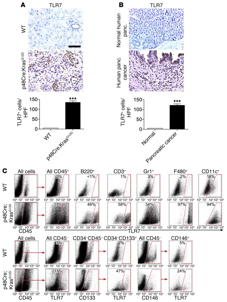
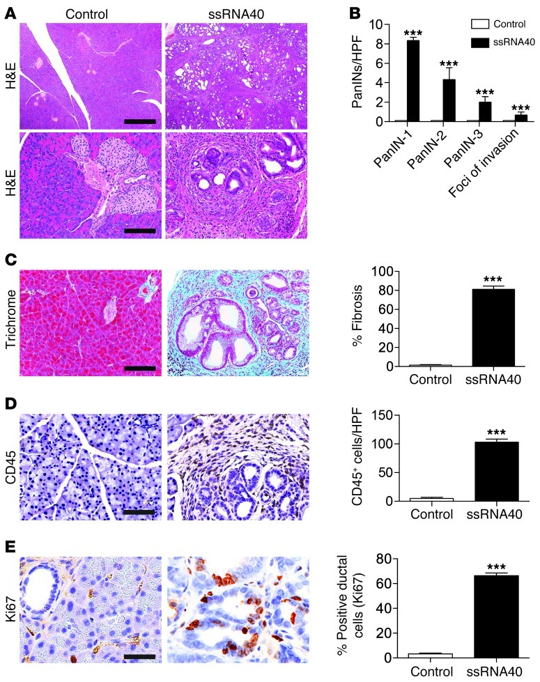
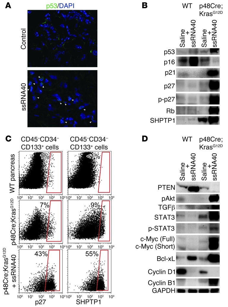
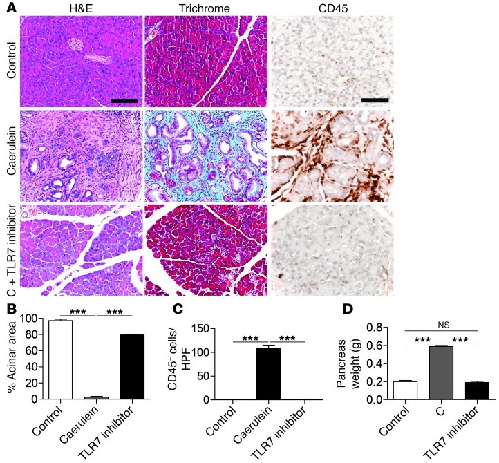
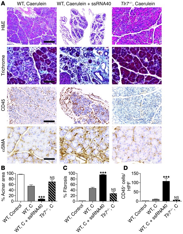
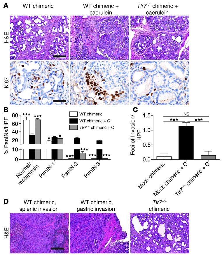
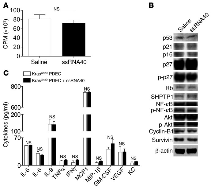
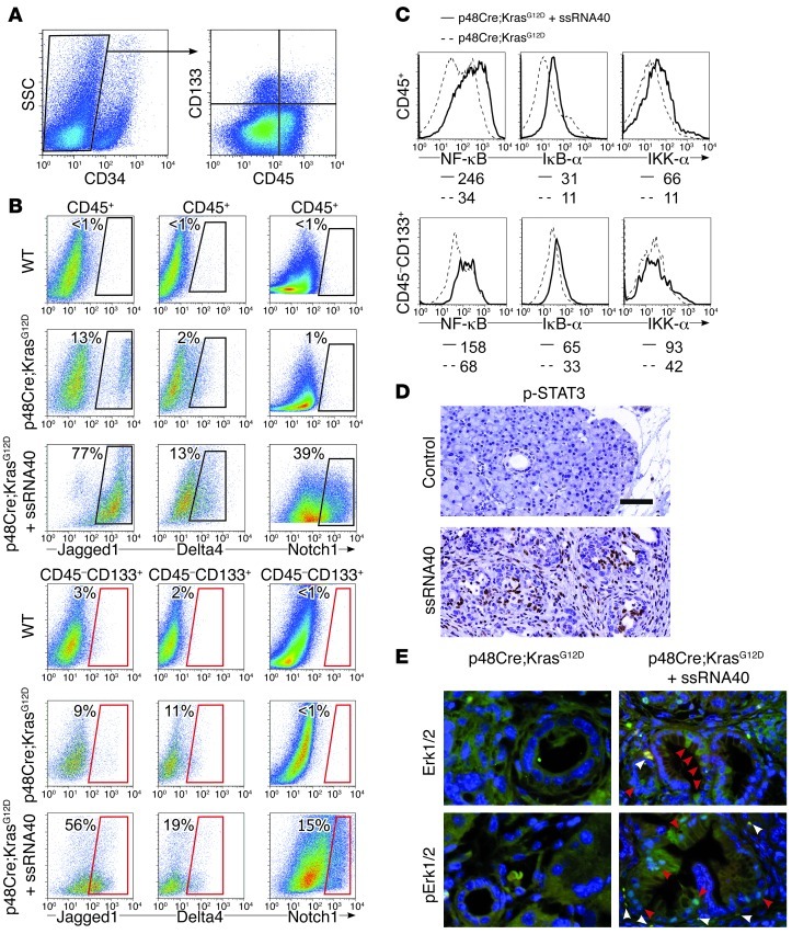
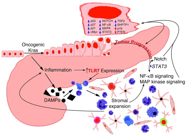
Pancreatic ductal adenocarcinoma is an aggressive cancer that interacts with stromal cells to produce a highly inflammatory tumor microenvironment that promotes tumor growth and invasiveness. The precise interplay between tumor and stroma remains poorly understood. TLRs mediate interactions between environmental stimuli and innate immunity and trigger proinflammatory signaling cascades. Our finding that TLR7 expression is upregulated in both epithelial and stromal compartments in human and murine pancreatic cancer led us to postulate that carcinogenesis is dependent on TLR7 signaling. In a mouse model of pancreatic cancer, TLR7 ligation vigorously accelerated tumor progression and induced loss of expression of PTEN, p16, and cyclin D1 and upregulation of p21, p27, p53, c-Myc, SHPTP1, TGF-β, PPARγ, and cyclin B1. Furthermore, TLR7 ligation induced STAT3 activation and interfaced with Notch as well as canonical NF-κB and MAP kinase pathways, but downregulated expression of Notch target genes. Moreover, blockade of TLR7 protected against carcinogenesis. Since pancreatic tumorigenesis requires stromal expansion, we proposed that TLR7 ligation modulates pancreatic cancer by driving stromal inflammation. Accordingly, we found that mice lacking TLR7 exclusively within their inflammatory cells were protected from neoplasia. These data suggest that targeting TLR7 holds promise for treatment of human pancreatic cancer.


Figures

Figure 1
Figure 1. High expression of TLR7 in inflammatory and epithelial cells in pancreatic carcinoma.
(A) Representative paraffin-embedded sections of pancreata from 6-month-old WT or p48Cre;KrasG12D mice. The number of TLR7+ cells per HPF was quantified (n = 6 per group). (B) Human normal (n = 5) and pancreatic cancer (n = 19) paraffin-embedded specimens were stained with mAbs directed against TLR7, and the number of TLR7+ cells per HPF was quantified. (C) Pancreatic leukocyte (CD45+) and (D) parenchymal (CD45) cellular subsets from 6-month-old WT and p48Cre;KrasG12D mice were analyzed by flow cytometry for expression of TLR7. The percentage of cells expressing TLR7 for each cellular subset is indicated. Data are representative of experiments repeated more than 3 times. Original magnification, ×40. Scale bar: 75 μm. ***P < 0.001.
Figure 2
Figure 2. TLR7 ligation accelerates pancreatic tumorigenesis.
6-week-old p48Cre;KrasG12D mice were administered saline or the TLR7 ligand ssRNA40 for 3 weeks. (A) Paraffin-embedded sections were examined by H&E. Original magnification, ×4 (top); ×20 (bottom). Scale bars: 700 μm (top); 150 μm (bottom). (B) The fractions of various grades of PanINs and foci of invasive cancer were quantified. (C) Gimori’s trichrome was performed to assess stromal fibrosis. Original magnification, ×20. Scale bar: 150 μm. (D) CD45 staining was performed to determine leukocytic infiltration. Original magnification, ×40. Scale bar: 75 μm. (E) Ki67 expression, a marker of proliferative index, was tested, and the percentage of Ki67+ ductal cells was determined by examining 10 HPFs per pancreas. Original magnification, ×80. Scale bar: 38 μm. n = 8 per group. ***P < 0.001.
Figure 3
Figure 3. TLR7 activation induces altered expression of tumor suppressor and oncogenic proteins.
6-week-old WT and p48Cre;KrasG12D mice were administered saline or ssRNA40 for 3 weeks. (A) Frozen sections of pancreata were stained with DAPI and an mAb directed against p53 and examined for colocalization (arrowheads) by confocal microscopy. Original magnification, ×63. (B) Western blots were performed on lysates using mAbs directed against p53, p16, p21, p27, p-p27, Rb, and SHPTP1. (C) CD45CD34CD133+ pancreatic epithelial cells were gated and analyzed for expression of p27 and SHPTP1 by flow cytometry. The percentage of positively staining cells for each respective marker is indicated. (D) Western blots were performed on lysates using antibodies directed against PTEN, pAKT, TGF-β, STAT3, p-STAT3, c-Myc, Bcl-xL, cyclin D1, cyclin B1, and GAPDH. Each Western blot was repeated at least 3 times with similar results.
Figure 4
Figure 4. Inhibition of TLR7 protects against pancreatic tumor progression.
6-week-old p48Cre;KrasG12D mice were treated with saline, caerulein (C), or caerulein plus an oligonucleotide inhibitor of TLR7. (A) Pancreata were assessed by H&E, trichrome, and CD45 staining. Original magnification, ×20 (H&E and trichrome); ×40 (CD45). Scale bars: 150 μm (H&E and trichrome); 75 μm (CD45). (B) Effacement of acini and (C) leukocytic infiltrate were quantified by examining 10 HPFs per mouse. (D) Whole pancreata were weighed. n = 8 per group. ***P < 0.001.
Figure 5
Figure 5. TLR7 ligation exacerbates pancreatic fibroinflammation, while TLR7 inhibition is protective.
Chronic pancreatitis was induced in 6-week-old WT or Tlr7–/– mice using caerulein. Selected cohorts of WT mice were additionally treated with ssRNA40. (A) Pancreata were stained using H&E, trichrome, or mAbs directed against CD45 and α-SMA. Original magnification, ×10 (H&E); ×20 (CD45); ×40 (trichrome and a-SMA). Scale bars: 300 μm (H&E); 150 μm (CD45); 75 μm (trichrome and a-SMA). (B) Surface area occupied by acinar units and (C) fibrotic surface area were determined using a computerized grid. (D) CD45+ leukocytic infiltrate was quantified by examining 10 HPFs per pancreas. n = 5 per group. ***P < 0.001.
Figure 6
Figure 6. TLR7 signaling in inflammatory cells regulates pancreatic cancer progression.
2-month-old p48Cre;KrasG12D mice were irradiated and made chimeric by bone marrow transfer from WT or Tlr7–/– mice. (A) 7 weeks later, mice were treated with either saline or caerulein to accelerate carcinogenesis. Mice were then sacrificed 3 weeks later, and pancreata were assessed by H&E and Ki67 staining. Original magnification, ×10 (H&E); ×60 (Ki67). Scale bars: 300 μm (H&E); 50 μm (Ki67). (B) The fraction of metaplastic and dysplastic ducts was measured for each cohort, and (C) the number of foci of invasive cancer was quantified using CK19 immunohistochemistry (n = 5 per group). *P < 0.05; ***P < 0.001. (D) Additional cohorts of chimeric mice (n = 4 per group) were not treated with caerulein, but kept until 12 months of life for histological analysis. Representative H&E-stained paraffin-embedded sections are shown. Original magnification, ×10. Scale bar: 300 μm.
Figure 7
Figure 7. TLR7 ligand does not directly activate PDECs.
(A) In vitro proliferation of p48Cre;KrasG12D PDECs was measured by uptake of [3H]-thymidine after stimulation with ssRNA40 or saline. (B) p48Cre;KrasG12D PDECs treated with ssRNA40 or saline were tested for expression of various tumor-suppressor or oncogenic proteins and for genes involved in regulation of apoptosis by Western blotting. (C) Cytokine production by p48Cre;KrasG12D PDECs was measured after stimulation with ssRNA40 or saline. Experiments were repeated 3 times and performed in triplicate.
Figure 8
Figure 8. TLR7 activation induces cell signaling changes in both peritumoral inflammatory and ductal epithelial cells in pancreatic cancer.
(A) To identify epithelial cells, CD34 pancreatic cellular suspensions from p48Cre;KrasG12D mice were gated and costained for CD133 and CD45. (B) CD45+or CD34CD45CD133+ pancreatic cells from 9-week-old WT mice or p48Cre;KrasG12D mice treated with saline or ssRNA40 for 3 weeks were analyzed for expression of Jagged1, Delta4, and Notch1 on flow cytometry. The percentage of positive cells for each respective marker is shown. Experiments were repeated twice using 2–4 mice per group with similar results. (C) CD45+ or CD34CD45CD133+ pancreatic cells from 9-week-old p48Cre;KrasG12D mice treated with saline or ssRNA40 were also analyzed for expression of NF-κB signaling intermediates. MFI is indicated. (D) Paraffin-embedded sections of saline and ssRNA40-treated p48Cre;KrasG12D mice were stained for p-STAT3. Representative images are shown. Original magnification, ×40. Scale bar: 75 μm. (E) Frozen sections of saline- and ssRNA40-treated p48Cre;KrasG12D mice were stained for DAPI and Erk1/2 or pErk1/2. Localization of Erk1/2 or pErk1/2 in inflammatory cells (white arrowhead) and ductal epithelial cells (red arrowhead) is indicated. Representative images are shown. Original magnification, ×40. Experiments were repeated twice.
Figure 9
Figure 9. Schematic depicting central position of TLR7 regulation of pancreatic tumorigenesis.

References

    1. Kuhn Y, Koscielny A, Glowka T, Hirner A, Kalff JC, Standop J. Postresection survival outcomes of pancreatic cancer according to demographic factors and socio-economic status. Eur J Surg Oncol. 2010;36(5):496–500. doi: 10.1016/j.ejso.2009.08.006. - DOI - PubMed
    1. Korc M. Pancreatic cancer-associated stroma production. Am J Surg. 2007;194(4 suppl):S84–S86. doi: 10.1016/j.amjsurg.2007.05.004. - DOI - PMC - PubMed
    1. Miyamoto H, Murakami T, Tsuchida K, Sugino H, Miyake H, Tashiro S. Tumor-stroma interaction of human pancreatic cancer: acquired resistance to anticancer drugs and proliferation regulation is dependent on extracellular matrix proteins. Pancreas. 2004;28(1):38–44. doi: 10.1097/00006676-200401000-00006. - DOI - PubMed
    1. De Wever O, Mareel M. Role of tissue stroma in cancer cell invasion. J Pathol. 2003;200(4):429–447. doi: 10.1002/path.1398. - DOI - PubMed
    1. Ding K, Lopez-Burks M, Sanchez-Duran JA, Korc M, Lander AD. Growth factor-induced shedding of syndecan-1 confers glypican-1 dependence on mitogenic responses of cancer cells. J Cell Biol. 2005;171(4):729–738. doi: 10.1083/jcb.200508010. - DOI - PMC - PubMed

Publication types

MeSH terms