. 2012 Dec 1;303(11):L967-77.
doi: 10.1152/ajplung.00144.2011. Epub 2012 Sep 28.

Stem cell conditioned medium improves acute lung injury in mice: in vivo evidence for stem cell paracrine action

Affiliations

Stem cell conditioned medium improves acute lung injury in mice: in vivo evidence for stem cell paracrine action

Lavinia Ionescu et al. Am J Physiol Lung Cell Mol Physiol. .

Abstract

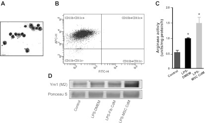
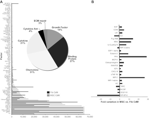
Mortality and morbidity of acute lung injury and acute respiratory distress syndrome remain high because of the lack of pharmacological therapies to prevent injury or promote repair. Mesenchymal stem cells (MSCs) prevent lung injury in various experimental models, despite a low proportion of donor-derived cell engraftment, suggesting that MSCs exert their beneficial effects via paracrine mechanisms. We hypothesized that soluble factors secreted by MSCs promote the resolution of lung injury in part by modulating alveolar macrophage (AM) function. We tested the therapeutic effect of MSC-derived conditioned medium (CdM) compared with whole MSCs, lung fibroblasts, and fibroblast-CdM. Intratracheal MSCs and MSC-CdM significantly attenuated lipopolysaccharide (LPS)-induced lung neutrophil influx, lung edema, and lung injury as assessed by an established lung injury score. MSC-CdM increased arginase-1 activity and Ym1 expression in LPS-exposed AMs. In vivo, AMs from LPS-MSC and LPS-MSC CdM lungs had enhanced expression of Ym1 and decreased expression of inducible nitric oxide synthase compared with untreated LPS mice. This suggests that MSC-CdM promotes alternative macrophage activation to an M2 "healer" phenotype. Comparative multiplex analysis of MSC- and fibroblast-CdM demonstrated that MSC-CdM contained several factors that may confer therapeutic benefit, including insulin-like growth factor I (IGF-I). Recombinant IGF-I partially reproduced the lung protective effect of MSC-CdM. In summary, MSCs act through a paracrine activity. MSC-CdM promotes the resolution of LPS-induced lung injury by attenuating lung inflammation and promoting a wound healing/anti-inflammatory M2 macrophage phenotype in part via IGF-I.


Figures

Fig. 1.
Fig. 1.
Characterization of mesenchymal stem cells (MSCs) isolated from C57BL/6 mice. A: MSCs differentiated along adipogenic, osteogenic, and chondrogenic lineages. Top: differentiated MSCs. Bottom: control MSCs. Left to right, respectively: oil red O staining (adipocytes), alizarin red (osteocytes), safranin O (chondrocytes). Size bar: 60 μm. B: representative flow-cytometry histograms. C: quantification of MSC surface marker expression. Values are expressed as means ± SE.
Fig. 2.
Fig. 2.
MSC-conditioned medium (CdM) decreased bronchoalveolar lavage fluid (BALF) total cell and neutrophil number in LPS-induced lung inflammation. A: LPS-DMEM mice (n = 10) had significantly more inflammatory cells than uninjured controls (n = 5) and control-CdM (n = 6) mice. Treatment with MSCs (n = 5) or MSC-CdM (n = 10), but not fibroblast (Fib; n = 5) or Fib-CdM (n = 5), significantly attenuated lung cells influx. BALF of mice treated with DMEM had 2 times more polymorphonuclear leukocytes (PMNs) than those treated with MSC-CdM. There were no differences in mononuclear cells (MN) number. *P < 0.05 control, control-CdM vs. LPS-MSC CdM; #P < 0.01 control, control-CdM vs. all other LPS groups (LPS-DMEM, LPS-Fib, LPS-Fib CdM, LPS-MSC); §P < 0.01 LPS-MSC vs. LPS-DMEM, LPS-Fib, LPS-Fib CdM. B: LPS-DMEM (n = 5) lungs had increased wet-to-dry weight ratios compared with control (n = 5) and control-CdM (n = 5) lungs. LPS-MSCs (n = 4) and LPS-MSC-CdM (n = 5), but not LPS-Fib (n = 4) or LPS-Fib-CdM (n = 5), had significantly decreased wet-to-dry weight ratios compared with LPS-DMEM. *P < 0.05 LPS-DMEM vs. LPS-MSC; †P < 0.05 LPS-Fib and LPS-Fib CdM vs. LPS-MSC CdM; #P < 0.01 control, control-CdM vs. LPS-DMEM, LPS-Fib, LPS-Fib CdM, LPS-MSC CdM; §P < 0.01 LPS-MSC vs. LPS-Fib and LPS-Fib CdM.
Fig. 3.
Fig. 3.
MSC-CdM reduced LPS-induced lung injury but failed to prevent body weight loss. A: no effect of treatment on LPS-induced body weight loss. Control (n = 13), control-CdM (n = 6), LPS-DMEM (n = 36), LPS-Fib (n = 9), LPS-Fib-CdM (n = 10), LPS-MSC (n = 8), LPS-MSC CdM (n = 35). **P < 0.01 control groups vs. each LPS group. B: LPS-MSC (n = 5) and LPS-MSC-CdM (n = 8) lungs had improved lung injury score compared with LPS-DMEM (n = 8), LPS-Fib (n = 5), and LPS-Fib CdM (n = 8). **P < 0.01 control groups vs. each LPS group; §P < 0.01 LPS-MSC and LPS-MSC CdM vs. LPS-DMEM, LPS-Fib, and LPS-Fib CdM. C: representative images of lungs from experimental animals. Size bar: 130 μm.
Fig. 4.
Fig. 4.
MSC-CdM promotes the M2 alveolar macrophage (AM) phenotype. A: representative photomicrograph of Hema3-stained AMs. Size bar: 15 μm. B: representative scatterplots of 4-color-stained AMs from experimental lungs. AMs were 98.2% CD11c+ and CD11b. FITC-H, fluorescein isothiocyanate; APC-H, allophycocyanin. C: AMs exposed to LPS for 24 h had greater arginase activity compared with control AMs. MSC-CdM enhanced arginase activity compared with DMEM (n = 4/group). *P < 0.05 control vs. LPS-DMEM and LPS-MSC-CdM; LPS-DMEM vs. LPS-MSC-CdM. D: immunoblots of AM show enhanced induced Ym1 expression in LPS-MSC-CdM compared with LPS-DMEM and LPS-Fib-CdM (n = 4 samples/lane).
Fig. 5.
Fig. 5.
MSC-CdM promotes the M2 AM phenotype in vivo. A: representative scatterplots of 4-color-stained AMs from experimental lungs. a.u., Arbitrary units. B: AMs from LPS-DMEM, LPS-Fib, LPS-Fib CdM lungs displayed an iNOS+Ym1 (gate P3; M1) phenotype. AMs from LPS-MSC and LPS-MSC CdM showed an iNOS-Ym1+ (gate P2; M2) phenotype (n = 5/group). Q1-1, Q1-2, Q1-3, Q1-4: quadrants resulted from quadrant gating. For M1 macrophages: no significant differences in control vs. control-CdM and LPS-MSC, control-CdM vs. LPS-MSC CdM, or LPS-DMEM vs. LPS-Fib CdM. P < 0.05 LPS-MSC vs. control-CdM and LPS-MSC CdM. P < 0.01 for all other group pairings. For M2 macrophages: no significant differences in control vs. LPS-Fib; LPS-DMEM vs. LPS-Fib CdM. P < 0.01 for all other group pairings.
Fig. 6.
Fig. 6.
A: MSC secretome analysis. Antibody array-based comparison between MSC-CdM and Fib-CdM. B: significant differences in MSC- vs. Fib-CdM levels of factors highlighted. Ant, antagonists.
Fig. 7.
Fig. 7.
IGF-I decreases LPS-induced lung injury. A: ELISA measurement showing that IGF-I is present at higher levels in MSC-CdM compared with Fib-CdM. *P < 0.05 control; #P < 0.01. BD: treatment with recombinant mouse IGF-I (rIGF-I; n = 5) significantly attenuated BALF cells influx (B), lung permeability (C), and lung injury score (D) compared with LPS-DMEM (n = 5). *P < 0.05 control vs. LPS groups; #P < 0.05 LPS-rIGF-I vs. LPS-DMEM. E: representative images of lungs from experimental animals. Size bar: 130 μm.
Fig. 8.
Fig. 8.
IGF-I promotes the M2 AM phenotype. A: representative scatterplots of 4-color-stained AMs from experimental lungs. B: LPS-rIGF-I treatment enhanced Ym1 expression and prevented inducible nitric oxide synthase (iNOS) induction compared with LPS-DMEM. LPS-Neut-CdM (LPS-MSC CdM +nAb IGF-I) attenuated Ym1 expression in AMs and was less effective in preventing iNOS induction (n = 5/group).

References

    1. Acquistapace A, Bru T, Lesault PF, Figeac F, Coudert AE, le Coz O, Christov C, Baudin X, Auber F, Yiou R, Dubois-Randé JL, Rodriguez AM. Human mesenchymal stem cells reprogram adult cardiomyocytes toward a progenitor-like state through partial cell fusion and mitochondria transfer. Stem Cells 29: 812–824, 2011 - PMC - PubMed
    1. Amariglio N, Hirshberg A, Scheithauer BW, Cohen Y, Loewenthal R, Trakhtenbrot L, Paz N, Koren-Michowitz M, Waldman D, Leider-Trejo L, Toren A, Constantini S, Rechavi G. Donor-derived brain tumor following neural stem cell transplantation in an ataxia telangiectasia patient. PLoS Med 6: e1000029, 2009 - PMC - PubMed
    1. Aslam M, Baveja R, Liang OD, Fernandez-Gonzalez A, Lee C, Mitsialis SA, Kourembanas S. Bone marrow stromal cells attenuate lung injury in a murine model of neonatal chronic lung disease. Am J Respir Crit Care Med 180: 1122–1130, 2009 - PMC - PubMed
    1. Bengatta S, Arnould C, Letavernier E, Monge M, de Preneuf HM, Werb Z, Ronco P, Lelongt B. MMP9 and SCF protect from apoptosis in acute kidney injury. J Am Soc Nephrol 20: 787–797, 2009 - PMC - PubMed
    1. Bonfield TL, Koloze M, Lennon DP, Zuchowski B, Yang SE, Caplan AI. Human mesenchymal stem cells suppress chronic airway inflammation in the murine ovalbumin asthma model. Am J Physiol Lung Cell Mol Physiol 299: L760–L770, 2010 - PMC - PubMed

Publication types

MeSH terms

Substances