. 2012 Dec;47(6):792-9.
doi: 10.1165/rcmb.2012-0145OC. Epub 2012 Sep 28.

Increased surfactant protein D fails to improve bacterial clearance and inflammation in serpinB1-/- mice

Affiliations

Increased surfactant protein D fails to improve bacterial clearance and inflammation in serpinB1-/- mice

J Michael Stolley et al. Am J Respir Cell Mol Biol. 2012 Dec.

Abstract

Previously, we described the protective role of the neutrophil serine protease inhibitor serpinB1 in preventing early mortality of Pseudomonas aeruginosa lung infection by fostering bacterial clearance and limiting inflammatory cytokines and proteolytic damage. Surfactant protein D (SP-D), which maintains the antiinflammatory pulmonary environment and mediates bacterial removal, was degraded in infected serpinB1-deficient mice. Based on the hypothesis that increased SP-D would rescue or mitigate the pathological effects of serpinB1 deletion, we generated two serpinB1(-/-) lines overexpressing lung-specific rat SP-D and inoculated the mice with P. aeruginosa. Contrary to predictions, bacterial counts in the lungs of SP-D(low)serpinB1(-/-) and SP-D(high) serpinB1(-/-) mice were 4 logs higher than wild-type and not different from serpinB1(-/-) mice. SP-D overexpression also failed to mitigate inflammation (TNF-α), lung injury (free protein, albumin), or excess neutrophil death (free myeloperoxidase, elastase). These pathological markers were higher for infected SP-D(high)serpinB1(-/-) mice than for serpinB1(-/-) mice, although the differences were not significant after controlling for multiple comparisons. The failure of transgenic SP-D to rescue antibacterial defense of serpinB1-deficient mice occurred despite 5-fold or 20-fold increased expression levels, largely normal structure, and dose-dependent bacteria-aggregating activity. SP-D of infected wild-type mice was intact in 43-kD monomers by reducing SDS-PAGE. By contrast, proteolytic fragments of 35, 17, and 8 kD were found in infected SP-D(low)serpinB1(-/-), SP-D(high) serpinB1(-/-) mice, and serpinB1(-/-) mice. Thus, although therapies to increase lung concentration of SP-D may have beneficial applications, the findings suggest that therapy with SP-D may not be beneficial for lung inflammation or infection if the underlying clinical condition includes excess proteolysis.


Figures
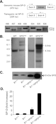
Figure 1.
Figure 1.
Generation of surfactant protein D (SP-D) transgenic mice and evaluation of SP-D expression. (A) PCR screening. Schematic showing the 228-bp band derived from rat cDNA and the endogenous 470-bp intron-spanning band. Wild-type (WT) mice (No. 496, 499, 504) produced only the larger band; transgenic mice (No. 497, 498, 505, and 506) produced both bands. The 228-bp transgenic band is more intense for No. 505 than for Nos. 497, 498, and 506, indicating higher transgene copy number. (B) Southern blot. Genomic DNA was digested with NcoI, and exon 7 of rat SP-D was used as probe. Tandemly integrated transgenes produced a 5.8-kb band and a unique band with size determined by location of the integration site; endogenous SP-D produces a 4.3-kb band. Copy number was estimated from the intensity ratio of the 5.8-kb band to the endogenous 4.3-kb band plus the number of integration sites. Shown from left are WT (no transgene), line SP-Dlow founded by No. 498 with two tandem transgene copies, WT, and SP-Dhigh founded by No. 505 with nine transgene copies and three integration sites. (C, D) SP-D in bronchoalveolar lavage fluid (BALF) of WT (serpinB1+/+, B1+/+); serpinB1−/− (B1−/−), transgenic SP-DlowserpinB1−/− (B1−/−SPDlow), and transgenic SP-DhighserpinB1−/− (B1−/−SPDhigh) mice examined by reducing SDS-PAGE and immunoblot. (C) Equal volumes of pooled BALF. (D) SP-D content determined by chemiluminescence of blots. BALF of individual mice was run undiluted (B1+/+, B1−/−) or diluted 1:3 (B1−/−SPDlow) or 1:5 (B1−/−SPDhigh) and band intensities were quantified by densitometry. Shown are means ± SEM for four mice per genotype.
Figure 2.
Figure 2.
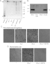
Structure–function characterization of SP-D in BALF of SP-D–overexpressing serpinB1−/− mice. (A) Native gel electrophoresis of pooled whole BALF of the four genotype mice diluted to approximately equal SP-D concentration. The immunoblot shows recombinant rat neck plus carbohydrate recognition domain (NCRD) (200 ng); recombinant rat SP-D (200 ng); BALF of WT mice (B1+/+); serpinB1−/− (B1−/−), SP-DlowserpinB1−/− (B1−/−SPDlow) (1:2 dilution), and SP-DhighserpinB1−/− (B1−/−SPDhigh) (1:8 and 1:16 dilution) mice; and recombinant human SP-D (200 ng). Samples were electrophoresed on 4 to 16% nativePAGE gels, transferred, and stained with CR-2011A antiserum; marker positions are indicated. Arrows on the left indicate SP-D dodecamers (lanes 2–8) and NCRD (lane 1); the dashed lines indicate the top of the gel. (B) Maltosyl affinity chromatography. Shown is an immunoblot of (left to right) pooled SP-DhighserpinB1−/− (B1−/−SPDhigh) BALF applied to the resin, nonadherent fraction, wash fraction, and EDTA eluate. Similar results were obtained for SP-DlowserpinB1−/− BALF (not shown). (C, D) Aggregation of Pseudomonas aeruginosa (C) and Escherichia coli (D) by the indicated volumes of maltosyl-purified SP-D of SP-DhighserpinB1−/− BALF. Shown are bright field images taken at 10× magnification.
Figure 3.
Figure 3.
P. aeruginosa lung infection of SP-D–overexpressing serpinB1−/− mice. Matched groups of WT (B1+/+), serpinB1−/− (B1−/−), SP-DlowserpinB1−/− mice (B1−/−SPDlow), and SP-DhighserpinB1−/− (B1−/−SPDhigh) mice were intranasally inoculated with 3 × 10⁶ colony-forming units (CFU) of strain PAO1. The mice were killed 24 hours later, and lung homogenates were prepared. (A) CFU per lung. Each symbol represents a value for an individual mouse; the horizontal lines show the medians, and the dotted line shows the limits of detection. (B) CFU per spleen determined for a (random) subset of mice. (C) TNF-α measured by ELISA. Means ± SEM are shown. (D) Immunoblots stained for SP-D of lung homogenates examined undiluted (B1+/+, B1−/−, and B1−/−SPDlow) or diluted 1 to 5 (B1−/−SPDhigh). Shown are representative samples from individual mice. Left lane: Std, recombinant rat SP-D. (AD) Data are from two experiments. (A, C) Differences among median or mean values were analyzed by Kruskal-Wallis ANOVA on Ranks followed by Dunn’s pairwise multiple comparisons.
Figure 4.
Figure 4.
Bronchoalveolar inflammation and injury of infected SP-D–overexpressing serpinB1−/− mice. WT (B1+/+), serpinB1−/− (B1−/−), and SP-D transgenic serpinB1−/− mice (B1−/−SPDlow and B1−/−SPDhigh) were intranasally inoculated with 107 CFU P. aeruginosa. BALF were prepared 24 hours later. (A) CFU per ml BALF. Symbols represent values for individual mice, and horizontal lines indicate medians. (B) Protein (left) and albumin (right). (C) Free elastase (left) and MPO (right). Data are from two experiments. (B, C) Means ± SEM are shown. (A C) Differences among median or mean values were analyzed by ANOVA followed by Holm-Sidak pairwise multiple comparisons except for albumin values, which were analyzed by Kruskal-Wallis ANOVA on Ranks followed by Dunn’s pairwise multiple comparisons.

References

    1. Cooley J, Takayama TK, Shapiro SD, Schechter NM, Remold-O’Donnell E. The serpin MNEI inhibits elastase-like and chymotrypsin-like serine proteases through efficient reactions at two active sites. Biochemistry 2001;40:15762–15770 - PubMed
    1. Fouret P, du Bois RM, Bernaudin JF, Takahashi H, Ferrans VJ, Crystal RG. Expression of the neutrophil elastase gene during human bone marrow cell differentiation. J Exp Med 1989;169:833–845 - PMC - PubMed
    1. Belaaouaj A, Kim KS, Shapiro SD. Degradation of outer membrane protein A in Escherichia coli killing by neutrophil elastase. Science 2000;289:1185–1188 - PubMed
    1. Reeves EP, Lu H, Jacobs HL, Messina CG, Bolsover S, Gabella G, Potma EO, Warley A, Roes J, Segal AW. Killing activity of neutrophils is mediated through activation of proteases by K+ flux. Nature 2002;416:291–297 - PubMed
    1. Moraes TJ, Zurawska JH, Downey GP. Neutrophil granule contents in the pathogenesis of lung injury. Curr Opin Hematol 2006;13:21–27 - PubMed

Publication types

MeSH terms