. 2012 Dec;7(12):1947-55.
doi: 10.2215/CJN.04800511. Epub 2012 Sep 27.

Differential diagnosis of lupus and primary membranous nephropathies by IgG subclass analysis

Affiliations

Differential diagnosis of lupus and primary membranous nephropathies by IgG subclass analysis

Young Soo Song et al. Clin J Am Soc Nephrol. 2012 Dec.

Abstract

Background and objectives: Previous studies showed that the accuracy of IgG subclasses (ISs) in differentiating membranous lupus nephritis (MLN) from primary membranous nephropathy (PMN) is <80%. This study hypothesized that diagnostic accuracy of ISs would be increased if renal compartment measurements and decision tree analysis are applied.

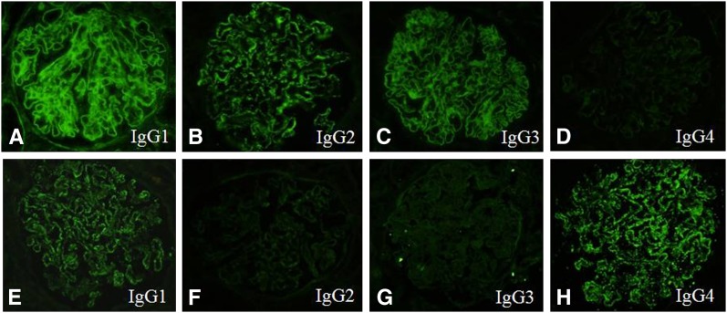
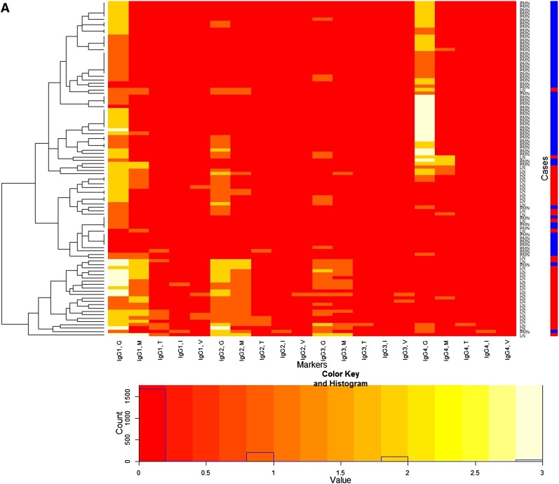
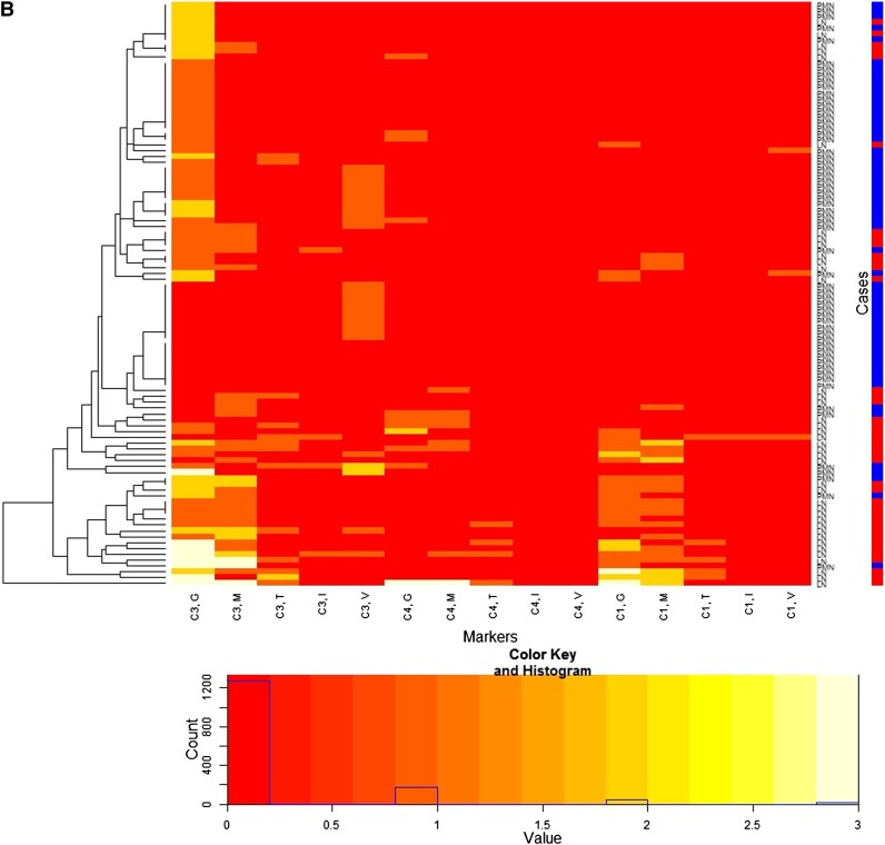
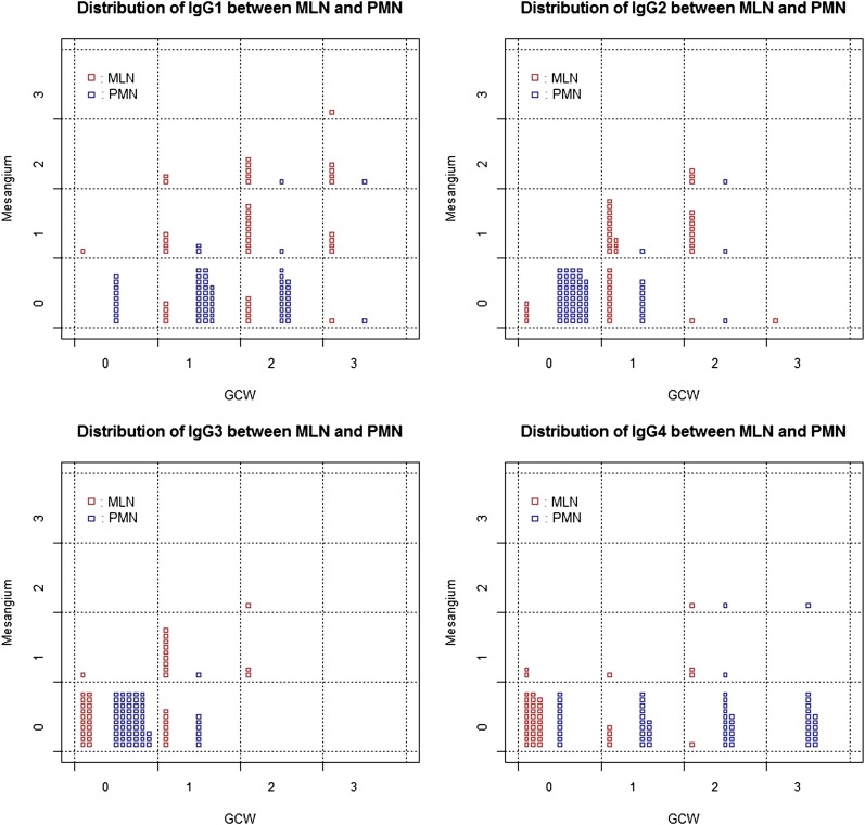
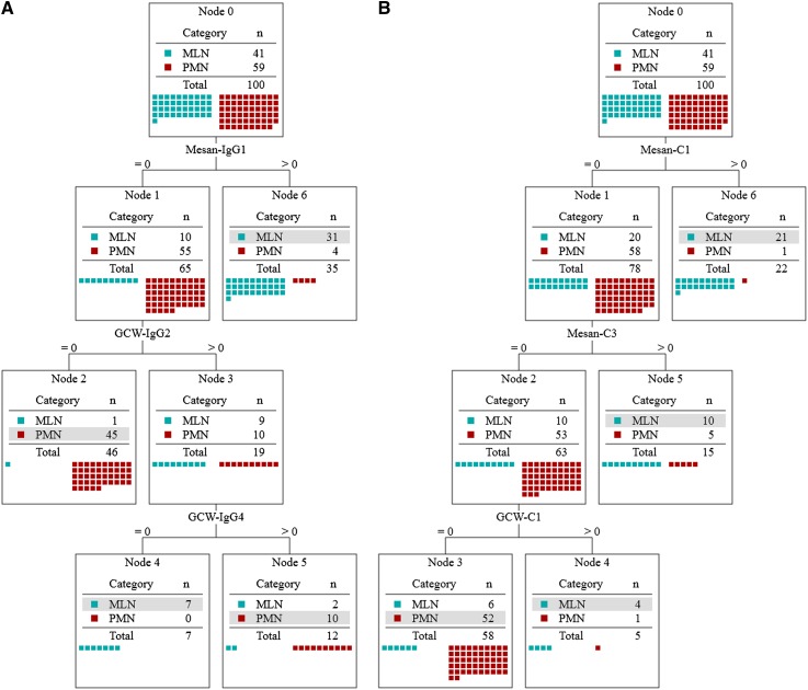
Design, setting, participants, & measurements: Renal biopsy specimens from 41 patients with MLN and 59 patients with PMN between October 2004 and March 2010 were examined, and immunofluorescence staining against IgG1, IgG2, IgG3, and IgG4 as well as C3, C1q, and C4 was evaluated in five different renal compartments (glomerular capillary walls, mesangium, tubules, interstitium, and blood vessels). From IS data, a decision tree to differentiate MLN from PMN was produced (IS decision tree) and its accuracy was compared with that of previous studies. Diagnostic accuracy of the IS decision tree was also compared with that of the complement decision tree as a reference.

Results: The demographic information and patterns of IS deposition were similar to those of previous studies. The IS decision tree had, as decision markers, IgG1 in the mesangium and IgG2 and IgG4 along the glomerular capillary wall. The IS decision tree showed higher accuracy (88%) than that of previous studies (<80%) and also that of the complement decision tree (81%).

Conclusions: Accuracy of ISs was increased due to the study methods, but the same methodology was less effective using complement measurements. Appropriate data analysis may enhance diagnostic value, but the analysis alone cannot achieve the ideal diagnostic value.


Figures

Figure 1.
Figure 1.
Immunofluorescence staining for IgG subclasses. A representative biopsy case of lupus nephritis, class V (A–D) and primary membranous nephropathy (E–H). Note the positive immunofluorescence staining for IgG1, IgG2, and IgG3 in the subepithelial portion along the glomerular capillary wall and in the mesangium (A–C) and the complete negative for IgG4 in the mesangium (D). A case of primary membranous nephropathy (E–H) shows strong immunofluorescence staining for IgG1 and IgG4 in the subepithelial portion along the glomerular capillary wall but negative in the mesangium.
Figure 2.
Figure 2.
Heatmap for immunofluorescence intensities on membranous lupus nephritis (LN) (41 patients) and primary membranous nephropathy (PMN) (59 patients) with a dendrogram (left side of the heatmap) and a color key and histogram (bottom of the heatmap) from IgG subclasses (A) and complements (B). The x-axis of the heatmap represents a list of markers and y-axis represents a list of patients, each of which is either LN or PMN. The patients are reordered and grouped according to the similarity of patients as shown in the dendrogram. Each cell represents an immunofluorescence intensity of the corresponding marker and patient, whose value can be 0, 1, 2, or 3 and its value is color coded from red through white as shown in the color key. The histogram shows counts of the cell values in the heatmap. Labels of rows are either LN or PMN, which is represented by the color bar along the label (LN is red, PMN is blue). The heatmap shows distinctive patterns of the overall distribution of the IgG subclasses for the patients with LN and PMN in the glomerular capillary wall (G), mesangium (M), tubular basement membrane (T), interstitium (I), and vessel (V). LN patients tend to show strong immunofluorescence staining for IgG1, IgG2, and IgG3 along the glomerular capillary wall and/or in the mesangium, whereas PMN patients tend to show strong immunofluorescence staining for IgG1 and IgG4 along the glomerular capillary wall. PMN patients are largely grouped into two.
Figure 2.
Figure 2.
Heatmap for immunofluorescence intensities on membranous lupus nephritis (LN) (41 patients) and primary membranous nephropathy (PMN) (59 patients) with a dendrogram (left side of the heatmap) and a color key and histogram (bottom of the heatmap) from IgG subclasses (A) and complements (B). The x-axis of the heatmap represents a list of markers and y-axis represents a list of patients, each of which is either LN or PMN. The patients are reordered and grouped according to the similarity of patients as shown in the dendrogram. Each cell represents an immunofluorescence intensity of the corresponding marker and patient, whose value can be 0, 1, 2, or 3 and its value is color coded from red through white as shown in the color key. The histogram shows counts of the cell values in the heatmap. Labels of rows are either LN or PMN, which is represented by the color bar along the label (LN is red, PMN is blue). The heatmap shows distinctive patterns of the overall distribution of the IgG subclasses for the patients with LN and PMN in the glomerular capillary wall (G), mesangium (M), tubular basement membrane (T), interstitium (I), and vessel (V). LN patients tend to show strong immunofluorescence staining for IgG1, IgG2, and IgG3 along the glomerular capillary wall and/or in the mesangium, whereas PMN patients tend to show strong immunofluorescence staining for IgG1 and IgG4 along the glomerular capillary wall. PMN patients are largely grouped into two.
Figure 3.
Figure 3.
Scattergrams with jittering showing the distribution of the immunofluorescence intensities of each IgG subclass both in the glomerular capillary wall (GCW) and mesangium between membranous lupus nephritis (MLN) and primary membranous nephropathy (PMN). The x-axes represent immunofluorescence intensities in GCW and the y-axes represent that in mesangium. A small rectangle in the cells represents a patient with either MLN (red) or PMN (blue) and the counts of the rectangles in each cell show the counts of the patients with the corresponding immunofluorescence value. MLN patients tend to show strong intensity for IgG1, IgG2, and IgG3 along the GCW and/or in the mesangium, whereas PMN patients tend to show strong intensity of IgG1 and IgG4 along the GCW.
Figure 4.
Figure 4.
Decision trees to differentiate membranous lupus nephritis (MLN) from primary membranous nephropathy (PMN) obtained from the profile of IgG subclass (A) and complements (B). Selected markers are the immunofluorescence intensities for IgG1 in the mesangium (Mesan) and IgG2 and IgG4 along the glomerular capillary wall (GCW) (A) (mean accuracy of 88.0% from 10-fold cross-validation; SEM 3.3%) and those for C1q and C3 in the mesangium and C1q along the GCW (mean accuracy of 81.0% from 10-fold cross-validation; SEM 3.8%). Starting from node 0, the path of decision is determined by the immunofluorescence value of each marker in the path and the class of a patient (MLN or PMN) is determined at a terminal node. Each node shows the distribution of classes; at each terminal node, the dominant class is shown as a gray box. The class of a patient is determined as a dominant class of a terminal node at which the patient arrived according to the decision path.

References

    1. Flanagan JG, Rabbitts TH: Arrangement of human immunoglobulin heavy chain constant region genes implies evolutionary duplication of a segment containing gamma, epsilon and alpha genes. Nature 300: 709–713, 1982 - PubMed
    1. Jefferis R: Polyclonal and monoclonal antibody reagents specific for IgG subclasses. Monogr Allergy 19: 71–85, 1986 - PubMed
    1. Hammarström L, Smith CI: IgG subclasses in bacterial infections. Monogr Allergy 19: 122–133, 1986 - PubMed
    1. Shakib F, Stanworth DR: Human IgG subclasses in health and disease. (A review). Part II. Ric Clin Lab 10: 561–580, 1980 - PubMed
    1. Shakib F, Stanworth DR: Human IgG subclasses in health and disease. (A review). Part I. Ric Clin Lab 10: 463–479, 1980 - PubMed

MeSH terms