Review
. 2013 Jan 1;3(1):a006551.
doi: 10.1101/cshperspect.a006551.

Molecular parallels between neural and vascular development

Affiliations
Review

Molecular parallels between neural and vascular development

Anne Eichmann et al. Cold Spring Harb Perspect Med. .

Abstract

The human central nervous system (CNS) features a network of ~400 miles of blood vessels that receives >20% of the body's cardiac output and uses most of its blood glucose. Many human diseases, including stroke, retinopathy, and cancer, are associated with the biology of CNS blood vessels. These vessels originate from extrinsic cell populations, including endothelial cells and pericytes that colonize the CNS and interact with glia and neurons to establish the blood-brain barrier and control cerebrovascular exchanges. Neurovascular interactions also play important roles in adult neurogenic niches, which harbor a unique population of neural stem cells that are intimately associated with blood vessels. We here review the cellular and molecular mechanisms required to establish the CNS vascular network, with a special focus on neurovascular interactions and the functions of vascular endothelial growth factors.


Figures

Figure 1.
Figure 1.
CNS vascularization. (A) Invasion of the spinal cord by ECs from the perineural vascular plexus. A quail neural tube (NT) section stained with QH1, an EC-specific monoclonal antibody. (Arrows) ECs from the (*) peri-neural vascular plexus (PNVP) invade the neural tissue on both sides of the floor plate (FP). (B) Schematic representation of EC invasion of the NT. Vessels from the PNVP invade the NT and grow toward the ventricle (V), where VEGF and Wnt factors are produced. (C) High magnification of the leading edge of capillaries invading the CNS. A leading tip cell extends filopodia toward hypoxic regions producing VEGF; note the opposing gradients of oxygen availability and VEGF production (triangles). Tip cells react to VEGF via VEGFR2 expressed on filopodia. Tip cell Dll4 activates Notch signaling on adjacent ECs, which differentiate into stalk cells that form a lumen allowing blood flow and tissue oxygenation. The PDGF-BB produced by tip cells promotes recruitment of pericytes expressing PDGFR-β.
Figure 2.
Figure 2.
Maturation of CNS blood vessels. (A) In brain capillaries, ECs form the BBB through formation of tight junctions and by limiting transport through cells by highly selective trancytosis (data not shown). The functional characteristics of this vasculature require interactions and continual cross talk between ECs, pericytes, astrocytes, and neural cells (data not shown). Only the astrocyte “end foot” (the tip of the cell that is physically connected to the capillary wall) is shown. The basal lamina, layers of extracellular matrix, surrounds the cells of the capillaries. (B) Confocal image of retinal tip cells stained with IsolectinB4 (red) and a closely associated NG2-positive pericyte (green). (C) Retinal ECs (red) also closely associate with GFAP-positive astrocytes. Magnification, 40× (B); 10× (C).
Figure 3.
Figure 3.
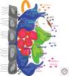
Interactions in the neurovascular niche. The niche is localized between the ependymal layer (E, gray) of lateral ventricles and a planar network of blood capillaries (bv, brown). It is composed of resident astroglial cells (type B, blue) including NSCs (type B1, blue), actively proliferating progenitors (type C, green), which generate neuroblasts (type A, red) migrating to the OB via the RMS. Quiescent NSCs are in subependymal position with an apical process in contact with the ventricular space, whereas actively proliferating progenitors preferentially associate with the surface of blood vessels. Both the ependyma and vessels express SDF1, which controls, through CXCR4, the homing of neural progenitors and NSCs in the SVZ and drives the expression of receptors that regulate proliferative behavior (EGFR) and cell adhesion (a6b1). Astroglial expression of the αv subfamily of integrins, including αvβ8, is critical for proper regulation of CNS angiogenesis and blood–brain barrier formation, by interaction with ECM molecules of the basement membrane, such as laminin. VEGFR family members have complementary expression pattern and function in the niche. The astroglial compartment expresses VEGFR-1 and VEGFR-3, the pool of neuroblasts expresses VEGFR-2, and ECs express VEGFR-1 and VEGFR-2. Coexpression of VEGFR-3 and EGFR in astrocytes characterizes NSCs. VEGF-C produced by SVZ astrocytes and also other cell types promotes activation of VEGFR-3-expressing cells, which increase in number following overexpression of VEGF-C. This enhances the number of type C cells and neuroblasts, without angiogenic effects in the niche. On the other hand, VEGF-A promotes angiogenesis and survival of neuroblasts. These effects are antagonized by astrocytes expressing VEGFR-1, which traps excess VEGF-A.

References

    1. Anderson KD, Pan L, Yang XM, Hughes VC, Walls JR, Dominguez MG, Simmons MV, Burfeind P, Xue Y, Wei Y, et al. 2011. Angiogenic sprouting into neural tissue requires Gpr124, an orphan G protein–coupled receptor. Proc Natl Acad Sci 108: 2807–2812 - PMC - PubMed
    1. Andreu-Agulló C, Morante-Redolat JM, Delgado AC, Fariñas I 2009. Vascular niche factor PEDF modulates Notch-dependent stemness in the adult subependymal zone. Nat Neurosci 12: 1514–1523 - PubMed
    1. Armulik A, Genové G, Mäe M, Nisancioglu MH, Wallgard E, Niaudet C, He L, Norlin J, Lindblom P, Strittmatter K, et al. 2010. Pericytes regulate the blood–brain barrier. Nature 468: 557–561 - PubMed
    1. Bautch VL, James JM 2009. Neurovascular development: The beginning of a beautiful friendship. Cell Adh Migr 3: 199–204 - PMC - PubMed
    1. Bellon A, Luchino J, Haigh K, Rougon G, Haigh J, Chauvet S, Mann F 2010. VEGFR2 (KDR/Flk1) signaling mediates axon growth in response to semaphorin 3E in the developing brain. Neuron 66: 205–219 - PubMed

MeSH terms

Substances

LinkOut - more resources