

Endosymbiotic calcifying bacteria: a new cue to the origin of calcification in metazoa?
- PMID: 23025593
- PMCID: PMC3485668
- DOI: 10.1111/j.1558-5646.2012.01676.x
Endosymbiotic calcifying bacteria: a new cue to the origin of calcification in metazoa?
Abstract
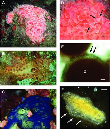
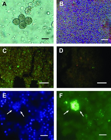
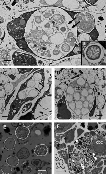
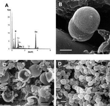
Sponges show the highest diversity of associated bacteria among marine invertebrates. Immunological evidence traces the origin of the sponge bacterial symbioses to the Precambrian era. Hence, sponges appear to be ideally suited for studying the evolutionary origins of prokaryote-metazoan associations. Sponges produce either calcareous or siliceous skeletons, which only coexist in a relict group of demosponges, the sclerosponges. We report here, for the first time, intensive calcification in nonsclerosponge siliceous demosponges. Calcification is mediated by endosymbiotic bacteria (calcibacteria) located in archeocyte-like sponge cells. These calcibacteria are devoid of bacterial walls and divide within sponge cells until they became surrounded by a calcitic sheet, being subsequently extruded to the sponge subectosomal (subepithelial) zone. Thousands of bacteria-produced calcitic spherules cover the surface of the host sponges, forming a cortex-like structure that mimics a rudimentary peripheral skeleton. Calcibacteria are vertically transferred to the sponge larvae during embryogenesis. Calcium detoxification may have generated this symbiotic association, with some additional benefits for the sponges, such as skeletal formation and deterrence from predation. This unique symbiosis holds implications for sponge biology and may advance discussions on the role of bacteria in early biocalcification processes in metazoans.
No Claim to original U.S. government works. Evolution© 2012 The Society for the Study of Evolution.
Figures
References
-
- Auguet JC, Casamayor EO. A hotspot for cold Crenarchaeota in the neuston of high mountain lakes. Environ. Microbiol. 2008;10:1080–1086. - PubMed
-
- Baumann P, Moran NA, Baumann L. Bacteriocyte-associated endosymbionts of insects. In: Dworkin M, editor. The prokaryotes. New York: Springer; 2000. pp. 1–67.
-
- Becerro MA, Turon X, Uriz MJ. Multiple functions for secondary metabolites in encrusting marine invertebrates. J. Chem. Ecol. 1997;23:1527–1547.
-
- Blanquer A, Uriz MJ, Agell G. Hidden diversity in sympatric sponges: adjusting life-history dynamics to share substrate. Mar. Ecol. Prog. Ser. 2008;371:109–115.
-
- Boury-Esnault N, Rützler K, editors. Smithsonian Contributions to Zoology 596. Washington, DC: Smithsonian Institution Press; 1997. Thesaurus of Sponge Morphology; pp. 1–55.
Publication types
MeSH terms
LinkOut - more resources
Full Text Sources
Other Literature Sources