. 2013 May 1;30(9):741-51.
doi: 10.1089/neu.2012.2478. Epub 2013 Feb 6.

Blast wave exposure impairs memory and decreases axon initial segment length

Affiliations

Blast wave exposure impairs memory and decreases axon initial segment length

Kelli L Baalman et al. J Neurotrauma. .

Abstract

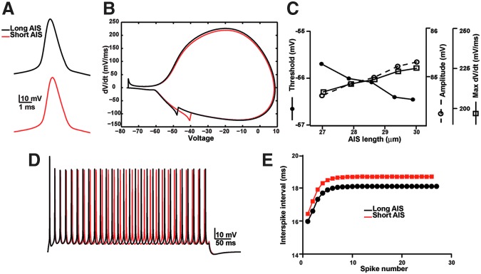
Exposure to a blast wave has been proposed to cause mild traumatic brain injury (mTBI), with symptoms including altered cognition, memory, and behavior. This idea, however, remains controversial, and the mechanisms of blast-induced brain injury remain unknown. To begin to resolve these questions, we constructed a simple compressed air shock tube, placed rats inside the tube, and exposed them to a highly reproducible and controlled blast wave. Consistent with the generation of a mild injury, 2 weeks after exposure to the blast, we found that motor performance was unaffected, and a panel of common injury markers showed little or no significant changes in expression in the cortex, corpus callosum, or hippocampus. Similarly, we were unable to detect elevated spectrin breakdown products in brains collected from blast-exposed rats. Using an object recognition task, however, we found that rats exposed to a blast wave spent significantly less time exploring a novel object when compared with control rats. Intriguingly, we also observed a significant shortening of the axon initial segment (AIS) in both the cortex and hippocampus of blast-exposed rats, suggesting altered neuronal excitability after exposure to a blast. A computational model showed that shortening the AIS increased both threshold and the interspike interval of repetitively firing neurons. These results support the conclusion that exposure to a single blast wave can lead to mTBI with accompanying cognitive impairment and subcellular changes in the molecular organization of neurons.


Figures

FIG. 1.
FIG. 1.
A compressed air-driven shock tube. (A) A photograph of the shock tube. Animals are positioned inside the end of the tube in a harness. (B) Schematic of the shock tube. (C) The average of the waveforms used in these experiments (the region in gray represents±standard error of the mean). (D) Three waveforms generated using different input pressures. (E) The amplitude of the overpressure scales linearly with the input pressure. (F) For a 1.25 m tube, the time-to-peak and to 1/2 max are relatively constant for the different input pressures used. (G) Frames from a high-speed video taken during a blast exposure. The original video was taken at 1000 frames/sec.
FIG. 2.
FIG. 2.
Exposure to a single blast wave does not cause changes in hemodynamics, weight gain, or motor behavior. (A-E) Mean blood pressure, systolic and diastolic blood pressure, tail blood flow, and tail blood volume were not significantly different in control animals compared with animals exposed to a blast immediately after blast exposure (post), 30 min after blast exposure (30' Post), or 1 h after blast exposure (60' Post). (F) Animals exposed to a single blast do not show any differences in weight at 48 h, 10 days, or 2 weeks after blast exposure. (G, H) Animals exposed to a single blast did not show impairment on the beam walking task (G, p>0.05) or the accelerating rotarod (H, p>0.05). Error bars indicate±standard error of the mean.
FIG. 3.
FIG. 3.
The expression levels of Iba1, glial fibrillary acidic protein (GFAP), and albumin are not significantly altered at 24 h or 2 weeks after blast exposure. (A-D) Immunostaining of cortex of blast-exposed and control rats using antibodies against Iba1 (A, C) and GFAP (B, D). (E-J) The ratio of immunofluorescence between blast-exposed and control rats in the cortex (CTX), corpus callosum (CC), and hippocampus (Hipp) 24 h and 2 weeks after blast exposure. After correcting for multiple comparisons, the only significant difference between blast and control animals was Iba1 immunoreactivity in the corpus callosum at 24 h after blast, and hippocampus at 2 weeks after blast. (K) The number of Iba1-labeled cells in hippocampus and cortex from control and blast-exposed rats at 24 hrs (5 blast-exposed and 4 control rats) and 2 weeks (7 blast-exposed and 4 control rats). (L) The number of Hoechst-labeled cells per field of view (FOV) in seven areas across the CTX from medial to lateral positions. Measurements were made at 2 weeks after blast-exposure (seven blast-exposed and four control rats). Error bars indicate±standard error of the mean. Scale bar=20 μm.
FIG. 4.
FIG. 4.
Immunoblot analysis of brains from blast-exposed rats shows no changes in the expression of injury markers 2 weeks after blast exposure. (A) Immunoblot analysis for PSD-95, βAPP, GFAP, and α-II spectrin. (B) Quantification of expression levels showed no significant differences between controls and animals exposed to a single blast. Actin and GAPDH were used as loading controls. Error bars indicate±standard error of the mean.
FIG. 5.
FIG. 5.
Exposure to a blast causes significant cognitive impairment and decrease in the length of the axon initial segment (AIS) in the hippocampus. (A, B) Schematic of the object recognition task. There was a 1-h interval between training and testing. (C) Control and blast-exposed rats spent equal amounts of time with both objects during the training phase. (D) Blast-exposed animals spent significantly less time with the novel object during testing compared with controls. (E) Immunostaining of the hippocampus using antibodies against βIV spectrin. The white box labels the area that was used for the length quantification. (F) Higher magnification image of βIV spectrin immunostaining in area CA1 of the hippocampus. The scale bar is 50 μm. (G) There is a significant decrease in the length of the AIS in injured animals compared with controls (p<0.05). Error bars indicate±standard error of the mean.
FIG. 6.
FIG. 6.
Exposure to a single blast wave causes a significant decrease in the length of the axon initial segment in the cortex. (A–H) Axon initial segment (AIS) were counted and the length was determined in two different locations in the cortex from seven blast-exposed and five control rats. Data from rostral regions are shown in A-D, while data from caudal regions are shown in E-H. A,B, E, F, The number of AIS in injured animals compared with controls was not significantly different from control animals (p>0.05 for βIV and ankG immunostaining). C, D, G, H, The lengths of the βIV spectrin and ankG-labeled AIS were significantly shorter in blast-exposed animals compared with controls (C, D, G, p<0.05 and H, p=0.08; two-way analysis of variance, significant main effect for blast exposure across all positions). A total of 14,078 and 11,575 AIS were measured from blast-exposed and control rats, respectively. (I) Immunoblot analysis of brain homogenates for AIS proteins shows no changes in the levels of AIS proteins or the generation of breakdown products in blast exposed animals. (J) Quantification of immunoblots revealed no significant changes in protein levels except for a decrease in ankB in blast exposed animals.
FIG. 7.
FIG. 7.
A computational model shows that a shorter axon initial segment (AIS) reduces neuronal excitability and alters the interspike interval. (A) Action potential shape for model neurons with AIS lengths of 30 μm (long AIS, black) or 28.65 μm (short AIS, red). (B) Phase plot of action potentials shown in A. (C) The amplitude of the action potential, threshold (defined as 20 mV/ms) and max dV/dt as a function of AIS length. (D) Voltage response of a 500 ms stimulation for neurons with AIS lengths of 30 μm (long AIS, black) or 28.65 μm (short AIS, red). (E) Interspike interval as a function of spike number for a model neuron with a long AIS (30 μm, black) or a short AIS (28.65 μm, red).

References

    1. Okie S. Traumatic brain injury in the war zone. N. Engl. J. Med. 2005;352:2043–2047. - PubMed
    1. Ling G.S. Ecklund J.M. Traumatic brain injury in modern war. Curr. Opin. Anaesthesiol. 2011;24:124–130. - PubMed
    1. Elsayed N.M. Toxicology of blast overpressure. Toxicology. 1997;121:1–15. - PubMed
    1. Mayorga M.A. The pathology of primary blast overpressure injury. Toxicology. 1997;121:17–28. - PubMed
    1. Leung L.Y. VandeVord P.J. Dal Cengio A.L. Bir C. Yang K.H. King A.I. Blast related neurotrauma: a review of cellular injury. Mol Cell Biomech. 2008;5:155–168. - PubMed

Publication types