Review
. 2013 Jan:100:30-47.
doi: 10.1016/j.pneurobio.2012.09.003. Epub 2012 Sep 29.

Emerging roles of Nrf2 and phase II antioxidant enzymes in neuroprotection

Affiliations
Review

Emerging roles of Nrf2 and phase II antioxidant enzymes in neuroprotection

Meijuan Zhang et al. Prog Neurobiol. 2013 Jan.

Abstract

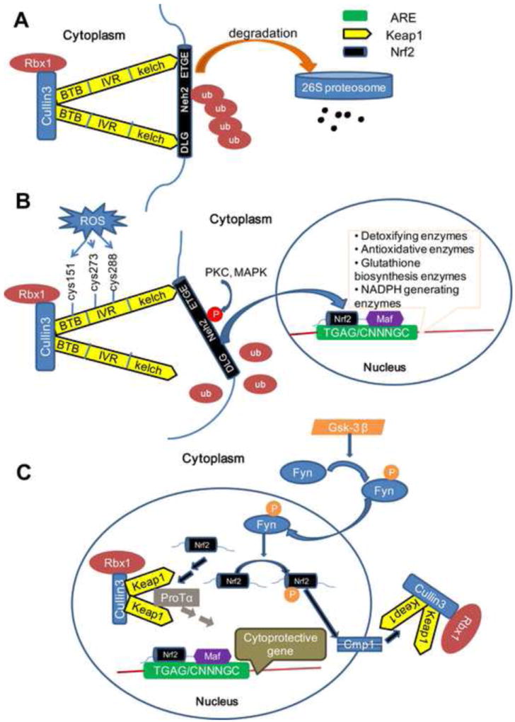
Phase II metabolic enzymes are a battery of critical proteins that detoxify xenobiotics by increasing their hydrophilicity and enhancing their disposal. These enzymes have long been studied for their preventative and protective effects against mutagens and carcinogens and for their regulation via the Keap1 (Kelch-like ECH associated protein 1)/Nrf2 (Nuclear factor erythroid 2 related factor 2)/ARE (antioxidant response elements) pathway. Recently, a series of studies have reported the altered expression of phase II genes in postmortem tissue of patients with various neurological diseases. These observations hint at a role for phase II enzymes in the evolution of such conditions. Furthermore, promising findings reveal that overexpression of phase II genes, either by genetic or chemical approaches, confers neuroprotection in vitro and in vivo. Therefore, there is a need to summarize the current literature on phase II genes in the central nervous system (CNS). This should help guide future studies on phase II genes as therapeutic targets in neurological diseases. In this review, we first briefly introduce the concept of phase I, II and III enzymes, with a special focus on phase II enzymes. We then discuss their expression regulation, their inducers and executors. Following this background, we expand our discussion to the neuroprotective effects of phase II enzymes and the potential application of Nrf2 inducers to the treatment of neurological diseases.


Figures

Figure 1
Figure 1. Classic model of Keap1/Nrf2/ARE signaling
(A) Under basal conditions, the Cul3-Keap1 complex sequesters Nrf2 in the cytosol by binding its ETGF and DLG motifs. This facilitates the ubiquitination and proteasomal degradation of Nrf2. (B) The DLG motif of Nrf2 is loosened from the Cul3-Keap1 complex when cells are exposed to ROS which blocks the ubiquitination and degradation of Nrf2. Following an intricate series of phosphorylations by several kinases, Nrf2 translocates into the nucleus and subsequently binds to the ARE elements by forming a heterodimer with Maf protein and initiating the transcription of phase II genes. (C) Nuclear Nrf2 can be phosphorylated by Fyn and be extruded back to the cytoplasm through the Cmp1 system. On the other hand, nuclear Nrf2 may also be sequestered by several Cul3-Keap1 complexes in the nucleus that are imported by ProTα. Both of these mechanisms help cells return back to basal conditions.
Figure 2
Figure 2. Structure of Nrf2: domain, phosphorylation sites and nuclear shuttling signals
Nrf2 has six domains. From N-terminus to C-terminus, these include Neh2, Neh4, Neh5, Neh6, Neh1 to Neh3. Four phosphorylation sites have been pinpointed - Ser40 in Heh2 domain by PKC, Ser342/Ser347 in Neh6 by GSK3! and Tyr 568 in Neh3 by Fyn. Three nuclear localization signals (NLS) have been identified - NLS1 located in Neh2, NLS2 located in Neh1, and NLS3 located in Neh 3. Two nuclear export sequences (NES) have also been identified - NES1 partially overlapped with Heh5, which contains a cysteine (Cys183) and NES2 located in Neh1. Purple line: phosphorylation sites; green box: NLS; red box: NES. CBP: CREB-binding protein; CHD6: chromo-ATPase/helicase DNA binding protein family member 6 (both CREB? and CHD6 are transcriptional co-activators); Maf: musculo-aponeurotic fibrosarcoma; TrCP: beta-transducin repeats-containing proteins, an E3 ligase.
Figure 3
Figure 3
Mechanisms of Nrf2 activation by different classes of inducers ( : Inducers : Ubiquitin) (A) Without inducers, Keap1 binds Nrf2 and facilitates the degradation of the Nrf2-Keap1 complex via Cul3. Under these circumstances, Nrf2 remains bound to Keap1 and is not free to translocate to the nucleus. (B) Inducers that act on the BTB domain. These interactions lead to structural changes of Keap1, blocking the binding of Cul3 to Keap1. Nrf2 is thereby protected from ubiquitinization. Consequently, the available pool of Nrf2 increases in size. (C) Inducers that act on IVR and interrupt the association of Keap1 with Nrf2. Translucent Nrf2 indicates that it cannot bind to Keap1. (D) Inducers that also act on IVR but interrupt the association of Keap1 with Cul3. (E) Inducers that act on the DGR domain and block the binding site of Nrf2. (F) Inducers that phosphorylate Nrf2.
Figure 4
Figure 4. The impact of Nrf2 on the antioxidant activities of Trx system
Trx can be oxidized to Trx-S2. This helps to reduce Protein-S2, H2O2 and Prx-S-S-Prx. Because Prx-SO3 cannot be reduced by Trx, Sfx restores Prx-SO3 back into the Trx cycle. Trx-S2 is reduced by TrxRs, with NADPH as the electron donor. The hydrogen and electron necessary for NADPH restoration come from pyruvate and the L-malate reaction cycle, which is catalyzed by the malic enzyme. The molecules that are subject to the control of Nrf2 are highlighted in red and include malic enzyme, TrxR-(SH)2,Trx-(SH)2, Prx-SH, Srxns. Abbreviations: GSH: glutathione; Prx: peroxiredoxin; Srxn: sulfiredoxin; Trx: thioredoxins; TrxR: thioredoxin reductase.
Figure 5
Figure 5. The impact of Nrf2 on the collaboration between neurons and astrocytes in GSH synthesis and function
GSH is synthesized from glutamate, cysteine and glycine, a reaction which is catalyzed by GCL and GS. Cysteine is the rate limiting substrate and GCL is the rate limiting biosynthetic enzyme in neurons. Cystine imported from xCT participates in astrocytic GSH synthesis. Synthesized GSH is then extruded by MRP1 and further cleaved by γGT and peptidases to generate free cysteine in the extracellular space. Cysteine then enters neurons through EAAC1 and facilitates neuronal GSH production and function. GSH exerts antioxidant effects by detoxifying H2O2, endogenous toxic and xenobiotic compounds and Pr-S-Pr. The molecules controlled by Nrf2 are highlighted in red. Abbreviations: Glu: glutamate; Cys: cysteine; Gly: glycine; γGluCys: γ-glutamylcysteine; CysGly: cysteinylglycine; GSSG: glutathione disulfide; GCL: γ-glutamylcysteine ligase; GS: glutathione synthase; Gpx: glutathione peroxidase; GR: glutathione reductase; GST: glutathione-S-transferase; γGT: γ-glutamyltransferase; Grx: glutaredoxin; Pr: protein; MRP1: multidrug resistance protein 1.

References

    1. Ahlgren-Beckendorf JA, Reising AM, Schander MA, Herdler JW, Johnson JA. Coordinate regulation of NAD(P)H:quinone oxidoreductase and glutathione-S-transferases in primary cultures of rat neurons and glia: role of the antioxidant/electrophile responsive element. Glia. 1999;25:131–142. - PubMed
    1. Alam J, Wicks C, Stewart D, Gong P, Touchard C, Otterbein S, Choi AM, Burow ME, Tou J. Mechanism of heme oxygenase-1 gene activation by cadmium in MCF-7 mammary epithelial cells. Role of p38 kinase and Nrf2 transcription factor. J Biol Chem. 2000;275:27694–27702. - PubMed
    1. Anderson MF, Blomstrand F, Blomstrand C, Eriksson PS, Nilsson M. Astrocytes and stroke: networking for survival? Neurochem Res. 2003;28:293–305. - PubMed
    1. Andoh T, Chock PB, Chiueh CC. The roles of thioredoxin in protection against oxidative stress-induced apoptosis in SH-SY5Y cells. J Biol Chem. 2002;277:9655–9660. - PubMed
    1. Aoyama K, Watabe M, Nakaki T. Regulation of neuronal glutathione synthesis. J Pharmacol Sci. 2008;108:227–238. - PubMed

Publication types

MeSH terms