

Brain tumors
- PMID: 23026359
- PMCID: PMC3925448
- DOI: 10.1053/j.semnuclmed.2012.06.001
Brain tumors
Abstract
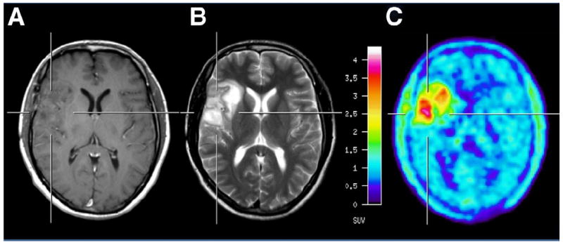
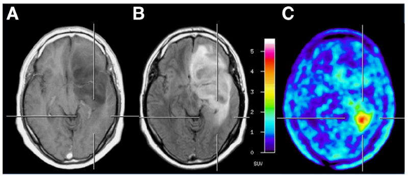
This review addresses the specific contributions of nuclear medicine techniques, and especially positron emission tomography (PET), for diagnosis and management of brain tumors. (18)F-Fluorodeoxyglucose PET has particular strengths in predicting prognosis and differentiating cerebral lymphoma from nonmalignant lesions. Amino acid tracers including (11)C-methionine, (18)F-fluoroethyltyrosine, and (18)F-L-3,4-dihydroxyphenylalanine provide high sensitivity, which is most useful for detecting recurrent or residual gliomas, including most low-grade gliomas. They also play an increasing role for planning and monitoring of therapy. (18)F-fluorothymidine can only be used in tumors with absent or broken blood-brain barrier and has potential for tumor grading and monitoring of therapy. Ligands for somatostatin receptors are of particular interest in pituitary adenomas and meningiomas. Tracers to image neovascularization, hypoxia, and phospholipid synthesis are under investigation for potential clinical use. All methods provide the maximum of information when used with image registration and fusion display with contrast-enhanced magnetic resonance imaging scans. Integration of PET and magnetic resonance imaging with stereotactic neuronavigation systems allows the targeting of stereotactic biopsies to obtain a more accurate histologic diagnosis and better planning of conformal and stereotactic radiotherapy.
Copyright © 2012 Elsevier Inc. All rights reserved.
Figures


References
-
- Kleihues P, Louis DN, Scheithauer BW, et al. The WHO classification of tumors of the nervous system. J Neuropathol Exp Neurol. 2002;61:215–225. discussion 226-219. - PubMed
-
- Reivich M, Kuhl D, Wolf A, et al. The [18F]fluorodeoxyglucose method for the measurement of local cerebral glucose utilization in man. Circ Res. 1979;44:127–137. - PubMed
-
- Hustinx R, Smith RJ, Benard F, et al. Can the standardized uptake value characterize primary brain tumors on FDG-PET? Eur J Nucl Med. 1999;26:1501–1509. - PubMed
-
- Spence AM, Muzi M, Mankoff DA, et al. 18F-FDG PET of gliomas at delayed intervals: Improved distinction between tumor and normal gray matter. J Nucl Med. 2004;45:1653–1659. - PubMed
-
- Prieto E, Martí-Climent JM, Domínguez-Prado I, et al. Voxel-based analysis of dual-time-point 18F-FDG PET images for brain tumor identification and delineation. J Nucl Med. 2011;52:865–872. - PubMed
Publication types
MeSH terms
Substances
Grants and funding
LinkOut - more resources
Full Text Sources
Medical
