

doi: 10.1007/978-1-62703-056-4_6.
A time-lapse imaging assay to study nuclear envelope breakdown
Affiliations
- PMID: 23027000
- PMCID: PMC4366132
- DOI: 10.1007/978-1-62703-056-4_6
Item in Clipboard
A time-lapse imaging assay to study nuclear envelope breakdown
Methods Mol Biol.
2013.
Abstract
Real-time imaging coupled with a permeabilized cell system presents a very versatile platform to visualize the dynamic and intricate nature of nuclear envelope breakdown, one of the major morphological changes of mitosis. Here, we describe such a strategy in which the plasma membrane of cells expressing fluorescently tagged nucleoporin POM121 and Histone H2B is permeabilized with digitonin. These cells are then incubated with mitotic Xenopus egg extract to create conditions that recapitulate the major events of mitotic nuclear remodeling seen in live-cell imaging, providing the opportunity to probe mechanisms and pathways that coordinate nuclear disassembly.
Figures

Live-imaging of nuclear envelope breakdown in intact cells. HeLa cells stably expressing POM121-3GFP were plated on a chambered cover glass slide. After ~16 h, the DMEM medium was aspirated and replaced with DMEM-F12 (which is buffered with HEPES and lacks phenol red) and the chamber was set up for live-imaging on a heated (37°C) automated microscope stage. DNA was visualized by adding DAPI, a cell permeable DNA dye, to the culture medium. Nuclear membrane remodeling and DNA condensation, two key features of early mitosis, were tracked to assess progression from interphase to prometaphase. Images were acquired using a ×60 objective in 3 min intervals. Scale bar, 10 μM.
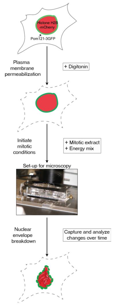
Experimental set-up for real-time nuclear envelope breakdown assay.HeLa cells stably expressing POM121-3GFP (green) and HistoneH2B-mCherry (red) are plated and allowed to grow on a chambered cover glass slide for 16–24 h (see Subheading 3.1). The cells are then treated with digitonin, which preferentially permeabilizes the plasma membrane, leaving the nuclear membrane intact (see Subheading 3.3). To initiate mitotic conditions, Xenopus mitotic extract and energy mix (see Subheading 3.2) are added to the permeabilized cells. The chambered cover glass slide is then placed in a slide holder and imaged as described in Subheading 3.4. As mitosis progresses, the mCherry-labeled chromatin condenses and POM121-3GFP tracks nuclear membrane remodeling.

Real-time imaging of nuclear envelope breakdown in permeabilized cells.HeLa cells stably expressing POM121-3GFP and Histone H2B-mCherry were permeabilized and incubated under mitotic (left panels) or interphase (right panels) conditions. Cells incubated with mitotic extract exhibited nuclear envelope breakdown and chromatin condensation characteristic of mitosis as time proceeded; arrows indicate when a particular nuclear envelope is first observed to lose continuity. The exact kinetics depends on the potency and quality of the batch of mitotic extract. In contrast, cells incubated with interphase extract maintain stable morphology over time, although the nuclei enlarge somewhat over time likely due to ongoing import. Scale bar, 20 μM.

Quantification of nuclear envelope breakdown in permeabilized cells The cumulative percentage of cells displaying broken/discontinuous POM121-3GFP-labeled nuclear envelope is plotted in a graph. Error bars indicate mean ± SD of four independent experiments in which >40 cells were counted in total.
References
-
- Guttinger S, Laurell E, Kutay U. Orchestrating nuclear envelope disassembly and reassembly during mitosis. Nat Rev Mol Cell Biol. 2009;10:178–191. - PubMed
-
- Cotter L, Allen TD, Kiseleva E, Goldberg MW. Nuclear membrane disassembly and rupture. J Mol Biol. 2007;369:683–695. - PubMed
-
- Georgatos SD, Pyrpasopoulou A, Theodoropoulos PA. Nuclear envelope breakdown in mammalian cells involves stepwise lamina disassembly and microtubule-drive deformation of the nuclear membrane. J Cell Sci. 1997;110(pt 17):2129–2140. - PubMed
-
- Beaudouin J, Gerlich D, Daigle N, Eils R, Ellenberg J. Nuclear envelope breakdown proceeds by microtubule-induced tearing of the lamina. Cell. 2002;108:83–96. - PubMed
Publication types
MeSH terms
Substances
Grants and funding
LinkOut - more resources
Full Text Sources
