

Neonatal seizures and status epilepticus
- PMID: 23027101
- PMCID: PMC3463810
- DOI: 10.1097/WNP.0b013e31826bd90d
Neonatal seizures and status epilepticus
Abstract
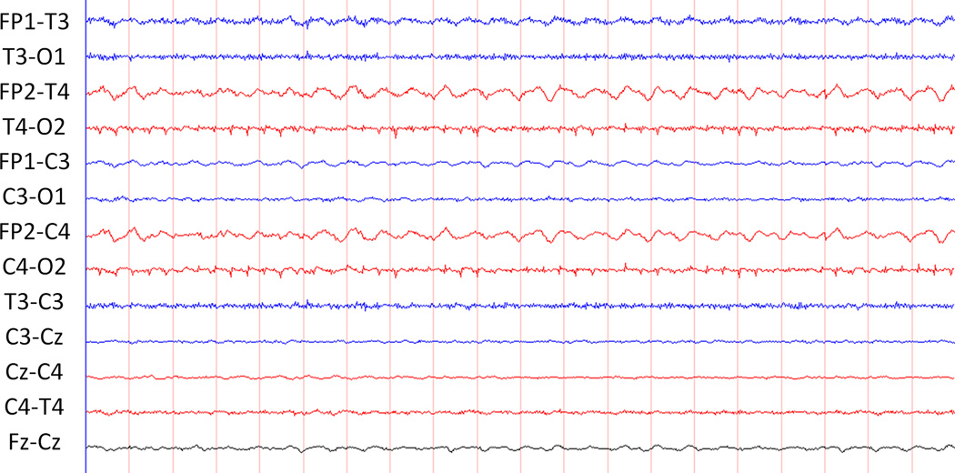
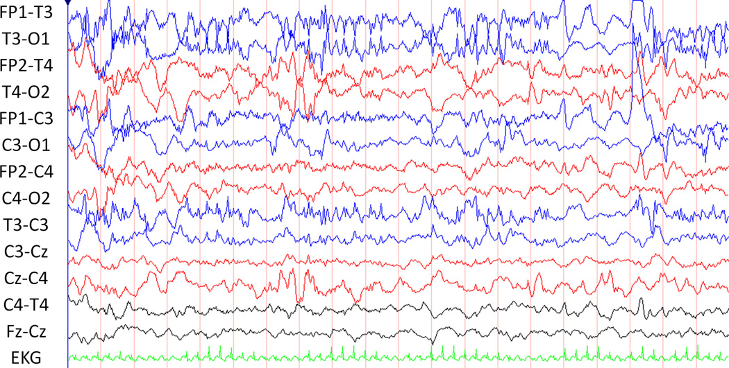
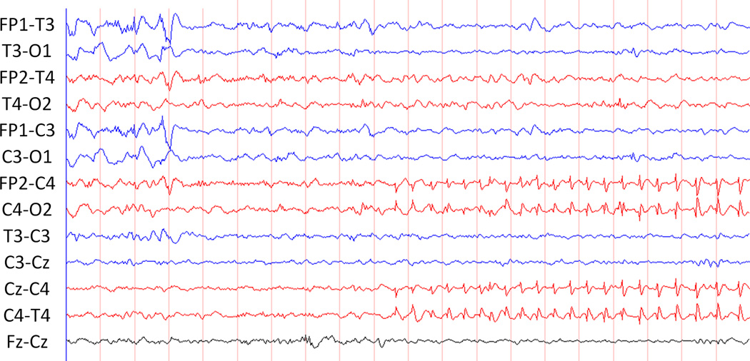
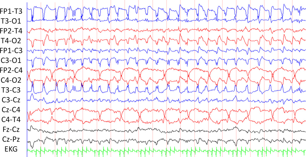
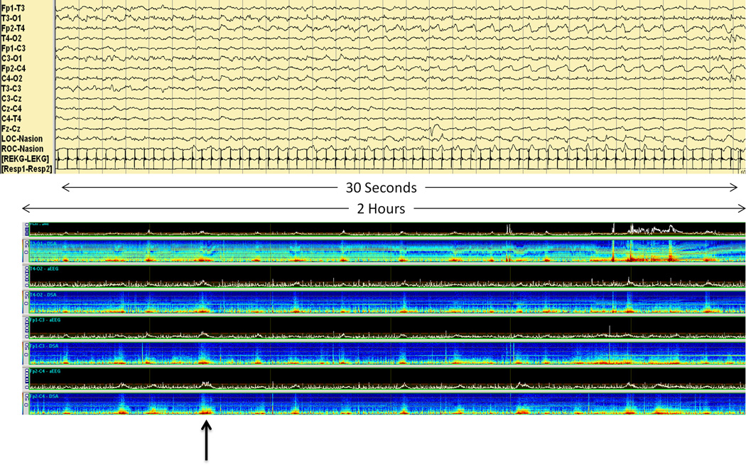
Neonatal seizures are common, often require EEG monitoring for diagnosis and management, may be associated with worse neurodevelopmental outcome, and can often be treated with existing anticonvulsants. A neonatal electrographic seizure is defined as a sudden, repetitive, evolving, and stereotyped event of abnormal electrographic pattern with amplitude of at least 2 μV and a minimum duration of 10 seconds. The diagnosis of neonatal seizures relies heavily on the neurophysiologist's interpretation of EEG. Consideration of specific criteria for the definition of a neonatal seizure, including seizure duration, location, morphology, evolution, semiology, and overall seizure burden, has utility for both the clinician and the researcher. The importance of EEG in the diagnosis and management of neonatal seizures, the electrographic characteristics of neonatal seizures, the impact of neonatal seizures on outcome, and tools to aid in the identification of neonatal seizures are reviewed.
Figures
References
-
- Abend NS, Dlugos D, Herman S. Neonatal seizure detection using multichannel display of envelope trend. Epilepsia. 2008;49:349–352. - PubMed
-
- Bellinger DC, Jonas RA, Rappaport LA, Wypij D, Wernovsky G, Kuban KC, Barnes PD, Holmes GL, Hickey PR, Strand RD, et al. Developmental and neurologic status of children after heart surgery with hypothermic circulatory arrest or low-flow cardiopulmonary bypass. N Engl J Med. 1995;332:549–555. - PubMed
-
- Berg A, Berkovic S, Brodie M, Buchhalter J, Cross J, Boas W, Engel J, French J, Glauser T, Mathern G, Moshe S, Nordli D, Plouin P, Scheffer I. Revised terminology and concepts for organization of seizures and epilepsies: Report of the ILAE Commission on Classification and Terminology 2005–2009. Epilepsia. 2010;51:676–685. - PubMed
-
- Bonifacio SL, Glass HC, Peloquin S, Ferriero DM. A new neurological focus in neonatal intensive care. Nat Rev Neurol. 2011;7:485–494. - PubMed
-
- Bourez-Swart MD, van Rooij L, Rizzo C, de Vries LS, Toet MC, Gebbink TA, Ezendam AG, van Huffelen AC. Detection of subclinical electroencephalographic seizure patterns with multichannel amplitude-integrated EEG in full-term neonates. Clin Neurophysiol. 2009;120:1916–1922. - PubMed
Publication types
MeSH terms
Substances
Grants and funding
LinkOut - more resources
Full Text Sources
Medical
