. 2012 Jul 24;56(3):e32.
doi: 10.4081/ejh.2012.e32.

Unusual localization and translocation of TRPV4 protein in cultured ventricular myocytes of the neonatal rat

Affiliations

Unusual localization and translocation of TRPV4 protein in cultured ventricular myocytes of the neonatal rat

Y Zhao et al. Eur J Histochem. .

Abstract

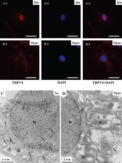
TRPV4 protein forms a Ca2+-permeable channel that is sensitive to osmotic and mechanical stimuli and responds to warm temperatures, and expresses widely in various kinds of tissues. As for cardiac myocytes, TRPV4 has been detected only at the mRNA level and there were few reports about subcellular localization of the protein. The purpose of the present study was to investigate the expression profile of TRPV4 protein in cultured neonatal rat ventricular myocytes. Using Western blots, immunofluorescence, confocal microscopy and immuno-electron microscopy, we have shown that TRPV4 protein was predominantly located in the nucleus of cultured neonatal myocytes. Furthermore, cardiac myocytes responded to hypotonic stimulation by translocating TRPV4 protein out of the nucleus. The significance and mechanism concerning the unusual distribution and translocation of TRPV4 protein in cardiac myocytes remain to be clarified.


Figures

Figure 1
Figure 1
Localization of TRPV4 protein in cardiac myocytes. A, B) Confocal images of freshly isolated (A1–3, scale bar: 15 µm) and cultured neonatal ventricular myocytes (B1–3, scale bar: 25 µm) labeled with anti-TRPV4 antibody (A1 and B1) and DAPI (A2 and B2), respectively, and the merged images (A3 and B3). The freshly isolated neonatal ventricular myocyte was small and round. C) Confocal images of freshly isolated adult rat ventricular myocytes labeled with anti-TRPV4 antibody (scale bar: 25 µm). D) Confocal image of the cultured ventricular myocytes in blank control (without TRPV4 antibody). E) Confocal image of the cultured ventricular myocytes in absorption test, in which the anti-TRPV4 antibody was preincubated with the peptide antigen. F, G) Immunolabeling TRPV4 protein in sections of the neonatal and adult ventricles.
Figure 2
Figure 2
Effects of hypotonicity on the distribution of TRPV4 in ventricular myocytes. Iso and Hypo: isotonic and hypotonic bath solutions, respectively. A, B) Immuno-localization of TRPV4 protein in cultured ventricular myocytes before (A) and after (B) hypotonic stimulation (scale bar: 25 µm). The myocytes were doubly labeled for TRPV4 protein (A-1, B-1) and the nucleus (A-2, B-2) as did as in Figure 1. A-3 and B-3 were correspondingly overlaid images. C, D) Immunoreactivity of TRPV4 protein detected by immuno-electron microscopy in cultured ventricular myocytes before (C) and after (D) hypotonic stimulation. N, nucleus; C, cytoplasm; arrows indicate the colloidal gold granules.
Figure 3
Figure 3
Hypotonically induced translocation of TRPV4 protein in cultured ventricular myocytes. A) The TRPV4 mRNA transcript was detected in adult renal tissues and cultured neonatal ventricular myocytes by RT-PCR amplification. B) Quantification of TRPV4 mRNA by real-time PCR for cultured ventricular myocytes in isotonic bath solution (Iso) and after hypotonic stimulation (Hypo). There were no significant differences at the mRNA levels between the two groups. C) Western blot analysis on the total TRPV4 protein of the freshly isolated adult ventricular myocytes and the corresponding absorption test. D) Western blot analysis on the total TRPV4 protein of cultured neonatal ventricular myocytes before and after exposure to hypotonic stimulation. E) Western blot analysis on TRPV4 protein in the nucleus fraction before and after hypotonic stimulation. F) Total and nuclear TRPV4 protein under isotonic and hypotonic conditions. The longitudinal coordinate stands for the relative ratio of TRPV4 fluorescent value contrast to β-actin fluorescent value (*P<0.05).

References

    1. Montell C, Rubin GM. Molecular characterization of the drosophila trp locus: A putative integral membrane protein required for phototransduction. Neuron. 1989;2:1313–23. - PubMed
    1. Koulen P, Cai Y, Geng L, Maeda Y, Nishimura S, Witzgall R, et al. Polycystin-2 is an intracellular calcium release channel. Nat Cell Biol. 2002;4:191–7. - PubMed
    1. Giamarchi A, Padilla F, Coste B, Raoux M, Crest M, Honoré E, et al. The versatile nature of the calcium-permeable cation channel TRPP2. EMBO Rep. 2006;7:787–93. - PMC - PubMed
    1. Loot AE, Popp R, Fisslthaler B, Vriens J, Nilius B, Fleming I. Role of cytochrome P450-dependent transient receptor potential V4 activation in flow-induced vasodi-latation. Cardiovasc Res. 2008;80:445–52. - PubMed
    1. Venkatachalam K, Hofmann T, Montell C. Lysosomal localization of TRPML3 depends on TRPML2 and the mucolipidosis-associated protein TRPML1. J Biol Chem. 2006;281:17517–27. - PMC - PubMed

Publication types