Review
. 2012 Aug 10;56(3):e38.
doi: 10.4081/ejh.2012.e38.

Ultrastructure of human mature oocytes after vitrification

Affiliations
Review

Ultrastructure of human mature oocytes after vitrification

M A Khalili et al. Eur J Histochem. .

Abstract

Since the introduction of human assisted reproduction, oocyte cryopreservation has been regarded as an attractive option to capitalize the reproductive potential of surplus oocytes and preserve female fertility. However, for two decades the endeavor to store oocytes has been limited by the not yet optimized methodologies, with the consequence of poor clinical outcome or of uncertain reproducibility. Vitrification has been developed as the promising technology of cryopreservation even if slow freezing remains a suitable choice. Nevertheless, the insufficiency of clinical and correlated multidisciplinary data is still stirring controversy on the impact of this technique on oocyte integrity. Morphological studies may actually provide a great insight in this debate. Phase contrast microscopy and other light microscopy techniques, including cytochemistry, provided substantial morphofunctional data on cryopreserved oocyte, but are unable to unraveling fine structural changes. The ultrastructural damage is one of the most adverse events associated with cryopreservation, as an effect of cryo-protectant toxicity, ice crystal formation and osmotic stress. Surprisingly, transmission electron microscopy has attracted only limited attention in the field of cryopreservation. In this review, the subcellular structure of human mature oocytes following vitrification is discussed at the light of most relevant ultrastructural studies.


Conflict of interest statement

Conflict of interests: all the authors declare there is no conflict of interest, personal, financial or otherwise, in relation of this work.

Figures

Figure 1
Figure 1
Fresh control (a, b), vitrified-warmed (c, d) and frozen-thawed by slow freezing (e, f) human oocytes. The general morphology and organelle microtopography are shown by LM (a, c, e) and TEM (b, d, f). No explicit differences in shape, dimensions and organelle distribution were found between fresh and cryopreserved oocytes. Apparent variations of ZP thickness (e) is an effect of the section plane (not equatorial). Vacuoles, detected only sporadically in both fresh (a, b) and vitrified-warmed (c, d) oocytes to the same extent, were numerous in the oocytes frozen-thawed by slow cooling (e, f). m, oocyte microvilli; O, oocyte; Va, vacuoles; ZP, zona pellucida; M-SER, mito-chondria-smooth endoplasmic reticulum aggregates. Scale bars: (a) 45 µm; (c, e) 40 µm; (b) 4 µm; (d, f) 5 µm. Panels a,b,e,f: modified from Coticchio et al.; panels c,d: modified from Nottola et al.
Figure 2
Figure 2
Vitrified-warmed (a) and frozen-thawed by slow freezing (b) human oocytes. TEM micrograph showing the paucity of vacuoles in (a) and the presence of an extensive vacuolization in (b). Va, vacuole; M, mitochondria. Scale bars: (a) 3 µm; (b) 2 µm. Panel b: modified from Coticchio et al.
Figure 3
Figure 3
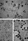
Fresh control (a) and vitrified-warmed (b, c) human oocytes. Mitochondria are generally rounded and provided with a few peripheral or transverse cristae. Dumb-bell shaped mitochondria (likely dividing) can be occasionally found in the ooplasm (a). Voluminous aggregates between mitochondria and smooth endoplasmic reticulum are seen (a, c). Note in (b) the absence of regularly shaped M-SER aggregates in the ooplasm. M, mitochondria; M-SER, mitochondria-smooth endoplasmic reticulum aggregates; M-V, mitochondria-vesicle complexes. Scale bars: (a) 2.5 µm; (b) 2 µm; (c) 0.5 µm. Modified from Nottola et al.
Figure 4
Figure 4
Fresh control (a) and vitrified-warmed (b, c) human oocytes. A rim of electron-dense CG (arrows) is seen just beneath the oolemma of the fresh oocyte (a). Instead, CG are sparse and form a discontinuous layer in vitrified-warmed oocytes (b, c). Note in (c) the increased compaction of the inner aspect of the ZP (asterisks) and the reduced number of microvilli. The dense ZP in c corresponds to an area of cortical cytoplasm virtually devoid of CG. CGs, cortical granules; m, microvilli; ZP, zona pellucida. Scale bars: (a) 1.1 µm; (b) 1.3 µm; (c) 2.5 µm. Panels a,b: modified from Nottola et al.

References

    1. Smith GD, Motta EE, Serafini P. Theoretical and experimental basis of oocyte vitrification. Reprod Biomed Online. 2011;23:298–306. - PubMed
    1. Gosden R. Cryopreservation: a cold look at technology for fertility preservation. Fertil Steril. 2011;96:264–8. - PubMed
    1. Cobo A, Diaz C. Clinical application of oocyte vitrification: a systematic review and meta-analysis of randomized controlled trials. Fertil Steril. 2011;96:277–85. - PubMed
    1. Tao T, Del Valle A. Human oocyte and ovarian tissue cryopreservation and its application. J Assist Reprod Genet. 2008;25:287–96. - PMC - PubMed
    1. Herrero L, Martínez M, Garcia-Velasco JA. Current status of human oocyte and embryo cryopreservation. Curr Opin Obstet Gynecol. 2011;23:245–50. - PubMed