. 2012 Dec;317(10):630-44.
doi: 10.1002/jez.1765. Epub 2012 Oct 1.

Localization of gonadotropin-releasing hormone (GnRH), gonadotropin-inhibitory hormone (GnIH), kisspeptin and GnRH receptor and their possible roles in testicular activities from birth to senescence in mice

Affiliations

Localization of gonadotropin-releasing hormone (GnRH), gonadotropin-inhibitory hormone (GnIH), kisspeptin and GnRH receptor and their possible roles in testicular activities from birth to senescence in mice

Shabana Anjum et al. J Exp Zool A Ecol Genet Physiol. 2012 Dec.

Abstract

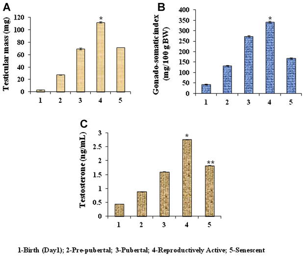
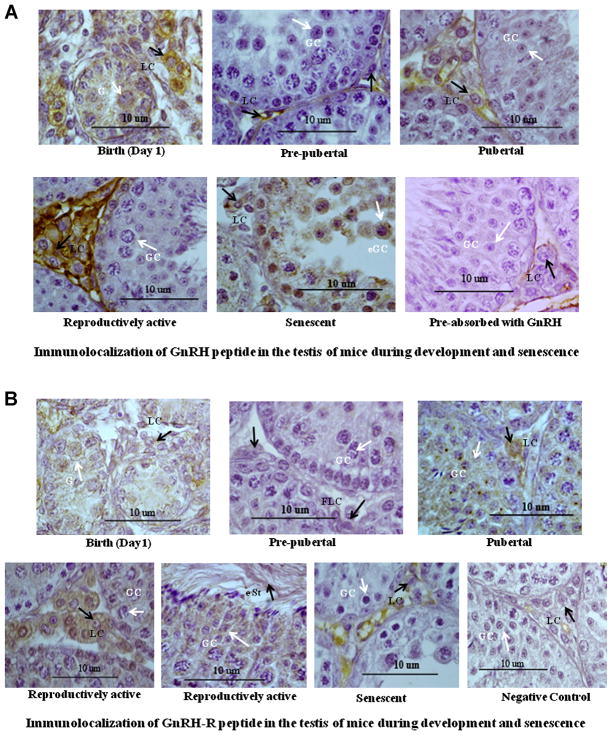
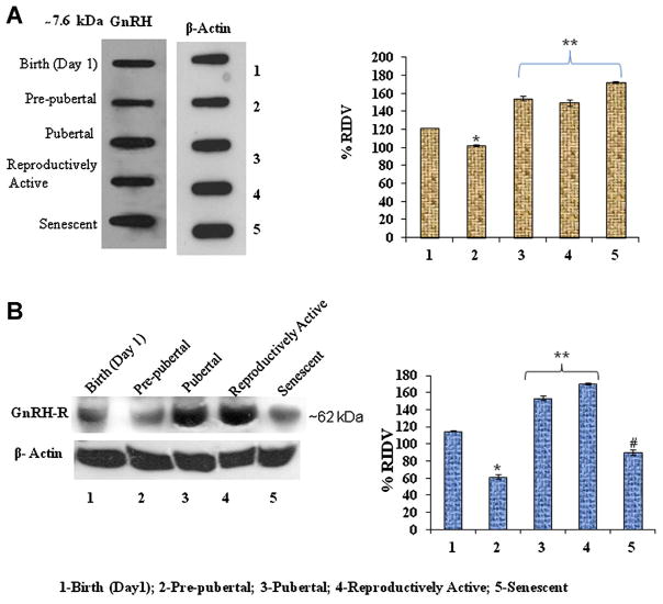
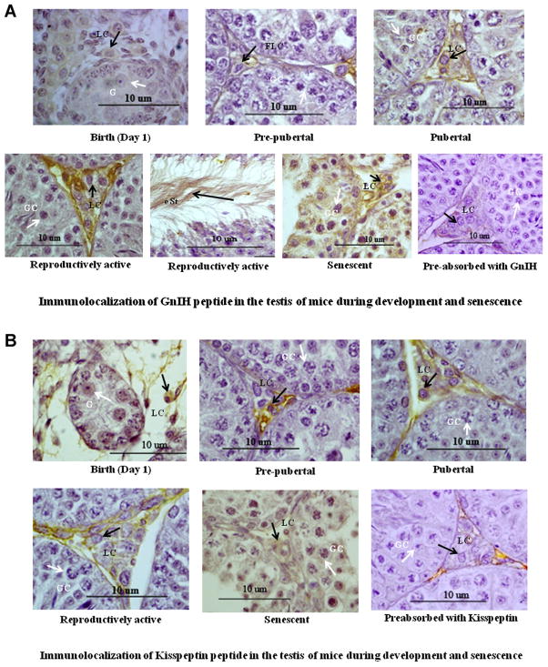
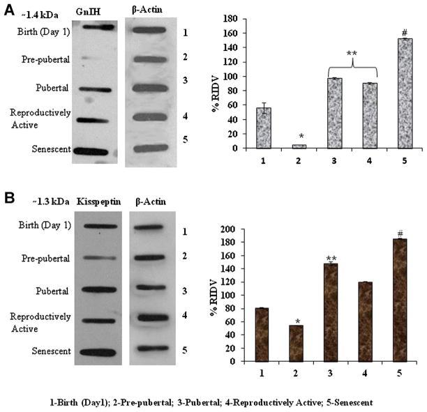
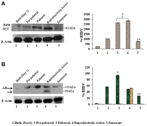
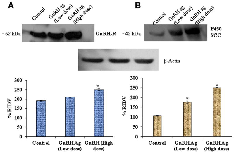
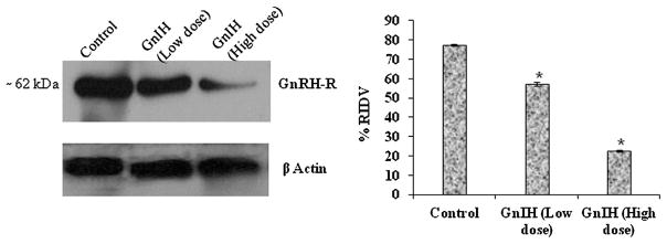
The changes in distribution and concentration of neuropeptides, gonadotropin-releasing hormone (GnRH), gonadotropin-inhibitory hormone (GnIH), kisspeptin, and gonadotropin-releasing hormone receptor (GnRH-R) were evaluated and compared with reproductive parameters, such as cytochrome P450 side-chain cleavage (P450 SCC) enzyme activity, androgen receptors (AR) in the testis and serum testosterone levels, from birth to senescence in mice. The results showed the localization of these molecules mainly in the interstitial and germ cells as well as showed significant variations in immunostatining from birth to senescence. It was found that increased staining of testicular GnRH-R coincided with increased steroidogenic activity during pubertal and adult stages, whereas decreased staining coincides with decreased steroidogenic activity during senescence. Similar changes in immunostaining were confirmed by Western/slot blot analysis. Thus, these results suggest a putative role of GnRH during testicular pubertal development and senescence. Treatment with a GnRH agonist ([DTrp(6), Pro(9)-NEt] GnRH) to mice from prepubertal to pubertal period showed a significant increase in steroidogenic activity of the mouse testis and provided further support to the role of GnRH in testicular pubertal maturation. The significant decline in GnRH-R during senescence may be due to a significant increase in GnIH synthesis during senescence causing the decrease in GnRH-R expression. It is considered that significant changes in the levels of GnRH-R may be responsible for changes in steroidogenesis that causes either pubertal activation or senescence in testis of mice. Furthermore, changes in the levels of GnRH-R may be modulated by interactions among GnRH, GnIH, and kisspeptin in the testis.


Figures

Figure 1
Figure 1
Age-dependent variation in (A) testicular mass and (B) gonadosomatic index (GSI), in mice. Values are represented as mean ± SEM (N = 8–10). *Values are significantly higher (P < 0.05) during reproductively active period vs. other age groups (birth, prepubertal, pubertal and senescent) of mice. C: Changes in serum testosterone levels in mice during development and senescence. Values are mean ± SEM. *Values are significantly higher during reproductively active period (P < 0.05) vs. birth, prepubertal, pubertal and senescence stages. **Values are significantly (P < 0.05) low during senescence versus reproductively active stage of mice. Birth (day 1); prepubertal (1 week); pubertal (6 weeks); reproductively active (15 weeks); and senescent (65 weeks).
Figure 2
Figure 2
A: Immunolocalization of GnRH peptide in the testis of mice during development and senescence. Immunoreactivity of GnRH increased in the interstitium during development. Strong immunostaining of GnRH was observed in the Leydig cell (LC) in the birth, pubertal, and reproductive active stages, whereas moderate immunostaining in germ cell (GC) of disorganized seminiferous tubule during senescence. Black arrow head showing immunostaining in Leydig cell (LC) whereas white arrow head showing immunostaining in gonocyte (G), germ cell (GC), and exfoliated germ cell (e GC) during birth and senescent, respectively. All figures are shown in 100 × magnification. B: Immunolocalization of GnRH receptor (GnRH-R) in the testis of mice during development and senescence. Immunoreactivity of GnRH-R increased in the interstitium during development. Strong immunostaining of GnRH-R was observed in the Leydig cell, germ cell as well as in elongated spermatids during pubertal and reproductive active stages. Mild immunostaining in Leydig cell and no immunoreactivity in germ cell in senescent. Black arrow head showing immunostaining in Leydig cell (LC) whereas white arrow head showing immunostaining in gonocyte (G), germ cell (GC) during birth, pubertal, reproductively active period and no immunostaining in germ cell (GC) of atrophied seminiferous tubule and senescent, respectively. All figures are shown in 100 × magnification. [Color figure can be seen in the online version of this article, available at http://onlinelibrary.wiley.com/journal/10.1002/(ISSN)1932-5231]
Figure 3
Figure 3
A: Slot blots analysis of GnRH peptide in the testis of Parkes strain mice of different age groups. *Values are significantly (P < 0.05) decreased in prepubertal period versus birth. **Values are significantly (P < 0.05) increased during puberty, reproductively active, and senescence versus birth and prepubertal stage of mice. B: Immunoblot analysis of GnRH-R protein expression in the testis of mice of different age groups. *Values are significantly (P < 0.05) decreased in prepubertal period versus birth. **Values are significantly (P < 0.05) increased during pubertal and reproductively active stages versus birth, prepubertal, and senescent mice. #Values are significantly (P < 0.05) decreased in senescence versus birth, pubertal, and reproductively active stages of mice. [Color figure can be seen in the online version of this article, available at http://onlinelibrary.wiley.com/journal/10.1002/(ISSN)1932-5231]
Figure 4
Figure 4
A: Immunolocalization of GnIH in the testis of mice during development and senescence. Immunoreactivity of GnIH showed mild staining in Leydig cell (LC) from birth to prepubertal stages, no staining in gonocyte (G) and strong staining in Leydig cell (LC) in the testis during pubertal and reproductively active period and in germ cells as well as in elongated spermatids during senescence of mice. Black arrow head showing immunostaining in Leydig cell (LC) whereas white arrow head showing immunostaining in germ cell (GC) during senescent. All figures are shown in 100 × magnification. B: Immunolocalization of kisspeptin in the testis of mice during development and senescence. Immunoreactivity of kisspeptin protein was mainly localized in the interstitium and Leydig cell in the testis. Immunoreactivity of kisspeptin showed mild to moderate staining in Leydig cell (LC) from Birth to prepubertal stages and strong staining in the Leydig cell (LC) during pubertal, reproductively active and in germ cells (GC) during senescent mice. Black arrow head showing immunostaining in Leydig cell (LC) whereas white arrow head showing immunostaining in germ cell (GC) during senescent. All figures are shown in 100 × magnification. [Color figure can be seen in the online version of this article, available at http://onlinelibrary.wiley.com/journal/10.1002/(ISSN)1932-5231]
Figure 5
Figure 5
A: Slot blots analysis of GnIH peptide expression in the testis of Parkes strain mice of different age groups. *Values are significantly decreased (P < 0.05) in prepubertal period vs. birth of mice. **Values are significantly increased (P < 0.05) during puberty, reproductively active vs. birth and prepubertal stage of mice. #Values are highly significant (P < 0.01) increased in senescence versus pubertal and reproductively active stage of mice. B: Slot blots analysis of kisspeptin protein expression in the testis of Parkes strain mice of different age groups. *Values are significantly decreased (P < 0.05) in prepubertal period versus birth of mice. **Values are significantly increased (P < 0.05) in pubertal versus reproductively active stage of mice. #Values are highly significant (P < 0.01) increased in senescence versus pubertal and reproductively active stage of mice. [Color figure can be seen in the online version of this article, available at http://onlinelibrary.wiley.com/journal/10.1002/(ISSN)1932-5231]
Figure 6
Figure 6
A: Immunoblot analysis of P450 SCC and AR expression in the testis of mice in different age groups. *Values are significantly increased (P < 0.05) during pubertal and reproductively active stages versus birth and prepubertal stage of mice. **Values are significantly declined (P < 0.05) in senescence versus prepubertal, pubertal, and reproductively active stage of mice. B: The immunoblot analysis of AR was detected two immunoreactive molecules against AR antibody in the reproductively active mice testes. *Values are significantly (P < 0.05) increased during pubertal stage versus birth, prepubertal, and reproductively active stages of mice. **Values are highly significant (P < 0.01) decreased in senescence versus prepubertal, pubertal, and reproductively active stages of mice. [Color figure can be seen in the online version of this article, available at http://onlinelibrary.wiley.com/journal/10.1002/(ISSN)1932-5231]
Figure 7
Figure 7
Effect of GnRH-agonist ([DTrp6, Pro9-NEt] GnRH) on GnRH-R and P450 SCC expressions. Different doses of GnRH treatment during prepubertal stage showed significant (P < 0.05) increased in expression of testicular GnRH-R in 2 μg/body weight versus control (A). *Values are significant (P < 0.05) increased in expression of testicular P450 SCC in both doses of GnRH (200 ng/day and 2 μg/day) versus control (B). [Color figure can be seen in the online version of this article, available at http://onlinelibrary.wiley.com/journal/10.1002/(ISSN)1932-5231]
Figure 8
Figure 8
Effect of GnIH on GnRH receptor (GnRH-R) protein expression in the testis. The in vitro study revealed that 1 and 10 ng/mL of GnIH significantly (P < 0.05) decreased the expression of testicular GnRH-R versus control. *Values are significant (P < 0.05) versus control.

References

    1. Adams TE. Using gonadotropin-releasing hormone (GnRH) and GnRH analogs to modulate testis function and enhance the productivity of domestic animals. Anim Reprod Sci. 2005;88:127–139. - PubMed
    1. Banerjee A, Singh A, Srivastava P, Turner H, Krishna A. Effects of chronic bhang (cannabis) administration on the reproductive system of male mice. Birth Defect Res B. 2011;92:195–205. - PubMed
    1. Bélanger A, Candas B, Dupont A, et al. Changes in serum concentrations of conjugated and unconjugated steroids in 40- to 80-year-old men. J Clin Endocrinol Metab. 1994;79:1086–1090. - PubMed
    1. Bhasin S, Heber D, Peterson M, Swerdloff R. Partial isolation and characterization of testicular GnRH-like factors. Endocrinology. 1983;112:1144–1146. - PubMed
    1. Botte MC, Chamagne AM, Carre MC, Counis R, Kottler ML. Fetal expression of GnRH and GnRH receptor genes in rat testis and ovary. J Endocrinol. 1998;159:179–189. - PubMed

Publication types