. 2012 Nov 16;287(47):40003-11.
doi: 10.1074/jbc.M112.382416. Epub 2012 Oct 1.

Interplay between menin and K-Ras in regulating lung adenocarcinoma

Affiliations

Interplay between menin and K-Ras in regulating lung adenocarcinoma

Yuan Wu et al. J Biol Chem. .

Abstract

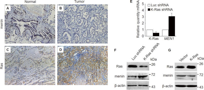
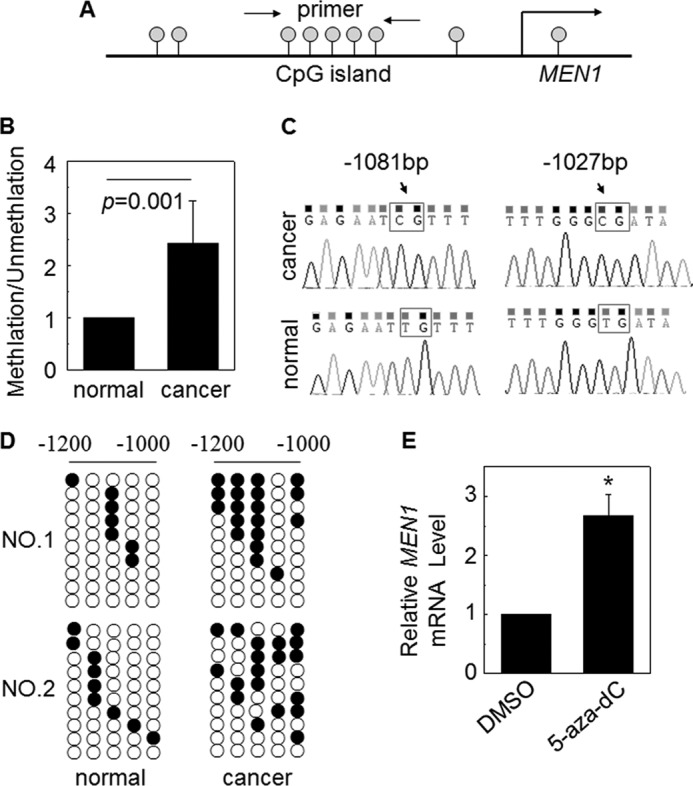
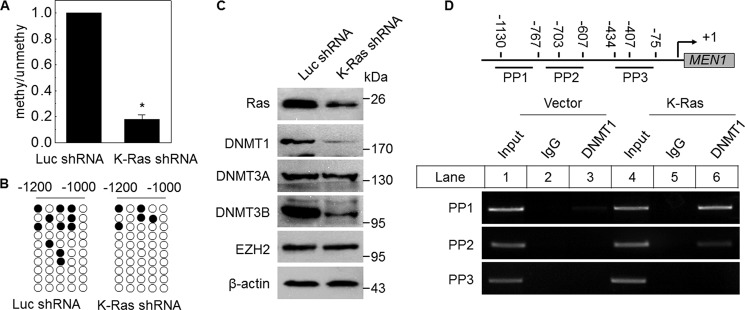
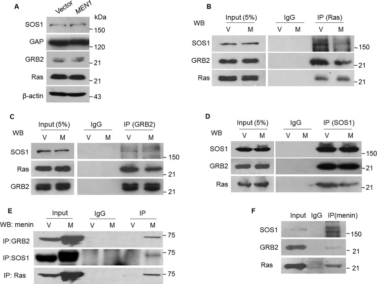
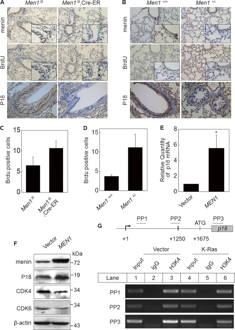
MEN1, which encodes the nuclear protein menin, acts as a tumor suppressor in lung cancer and is often inactivated in human primary lung adenocarcinoma. Here, we show that the inactivation of MEN1 is associated with increased DNA methylation at the MEN1 promoter by K-Ras. On one hand, the activated K-Ras up-regulates the expression of DNA methyltransferases and enhances the binding of DNA methyltransferase 1 to the MEN1 promoter, leading to increased DNA methylation at the MEN1 gene in lung cancer cells; on the other hand, menin reduces the level of active Ras-GTP at least partly by preventing GRB2 and SOS1 from binding to Ras, without affecting the expression of GRB2 and SOS1. In human lung adenocarcinoma samples, we further demonstrate that reduced menin expression is associated with the enhanced expression of Ras (p < 0.05). Finally, excision of the Men1 gene markedly accelerates the K-Ras(G12D)-induced tumor formation in the Men1(f/f);K-Ras(G12D/+);Cre ER mouse model. Together, these findings uncover a previously unknown link between activated K-Ras and menin, an important interplay governing tumor activation and suppression in the development of lung cancer.


Figures

FIGURE 1.
FIGURE 1.
Down-regulated menin expression was correlated with up-regulated Ras. A–D, sections from paraffin-embedded adjacent normal tissues and cancer tissues were stained with antibodies against menin (A and B) and Ras (C and D) for IHC staining, respectively, Original magnification, ×200. E and F, the expression of menin, detected by real time qRT-PCR (E) and Western blot (F), was increased in A549 cells that were transfected with shRNA vector against K-Ras compared with luciferase. G, menin expression, detected by Western blot, was reduced in K-Ras overexpression A549 cells.
FIGURE 2.
FIGURE 2.
Down-regulated menin expression was partly associated with promoter DNA methylation. A, the scheme shows the CpG islands of MEN1 promoter that is the aim of the MS-PCR primer. B, MS-PCR analysis of the MEN1 promoter methylation of lung cancer tissue and adjacent normal tissue, obtained from the surgeries of six patients. The methylation/unmethylation of normal samples was set to 1 (n = 6). C, the representative bisulfite sequencing chromatograms of cancer and normal tissues in the two MEN1 promoter CpG sites. D, DNA methylation analysis by bisulfite cloning and sequencing. The analysis was carried out for two paired cancer and normal tissues bisulfite sequencing. Each circle in each row represents a single CpG site. Each line represents an individually sequenced clone, and circles represent CpG residues. White and black circles represent unmethylated and methylated CpG sites, respectively. E, A549 cells were either treated or not treated with DNA methylation inhibitor 5-aza-dc (5 μm). Real time qRT-PCR was performed to detect the MEN1 mRNA expression. *, p < 0.05 versus control. DMSO, dimethyl sulfoxide.
FIGURE 3.
FIGURE 3.
K-Ras increases the MEN1 promoter DNA methylation via DNMTs. A, quantitative MS-PCR was used to analyze the methylation level of MEN1 promoter in K-Ras knockdown A549 cells (K-Ras shRNA), compared with Luc shRNA control. *, p < 0.05 versus control. B, bisulfite sequence analysis of each clone from K-Ras shRNA or Luc shRNA A549 cells (10 clones/sample). C, Western bolt detection of DNMTs and EZH2 in K-Ras diminished A549 cells. D, a schematic representation of the MEN1 gene loci and amplicons used for ChIP assay. ChIP assay was performed using the antibody against DNMT1 in vector and K-Ras-transfected A549 cells. PCR was carried out using primers for each amplicon.
FIGURE 4.
FIGURE 4.
Menin suppresses Ras GTP activity. A, Western blotting detected the impact of menin overexpression on SOS1, GAP, GRB2, and Ras expression in A549 cells. B–E, menin-overexpressed and control A549 cells lysates were immunoprecipitated for Ras, GRB2, or SOS1, and the immunoprecipitates were analyzed by Western blotting (WB) for the presence of SOS1, Ras, GRB2, and menin. F, total protein lysates of A549 cells were used for endogenous immunoprecipitation with antibody to menin and IgG as a control, followed by Western blotting for SOS1, GRB2, and Ras. V, vector; M, MEN1.
FIGURE 5.
FIGURE 5.
Men1 KO promotes lung epithelial cell proliferation. A, control Men1l/l and Men1l/l;Cre-ER mice (n = 8 mice) were fed tamoxifen at the age of 12 weeks at 200 mg/kg of body weight/day for 2 consecutive days. Four weeks after the last dose of tamoxifen feeding, the mice were sacrificed. The lung tissue sections from Men1l/l and Men1l/l;Cre-ER mice were stained with antibody of menin, BrdU, and p18. B, sections from paraffin-embedded Men+/+ and Men1+/− mice lung (3 months old) samples were also immunostained with menin, BrdU, and p18 antibody (-200; insets, ×400). C and D, quantification of BrdU+ lung cells from the groups of Men1l/l (control) versus tamoxifen-fed Men1l/l;Cre-ER mice and Men +/+ (control) versus Men1+/− mice. *, p < 0.05 versus control. E, real time qRT-PCR shows p18 mRNA expression in vector- or menin-expressing A549 cells. F, Western blot analysis the expression of P18, CDK4, and CDK6 in vector and menin overexpression A549 cells. G, a schematic representation of the p18 gene loci and amplicons used for ChIP assay. ChIP assay using the antibody against H3K4me3 in vector and K-Ras transfected A549 cells.
FIGURE 6.
FIGURE 6.
Menin excision promotes the development of K-Ras-induced lung cancer. A, the lung tissue hematoxylin and eosin staining of K-RasG12D/+ mice (C57BL/6 and 129/Sv mixed background) and Men1f/f;K-RasG12D/+ mice (K-RasG12D/+ mice crossed with Men1f/f mice) fed tamoxifen. B and C, the quantitative analysis of the number and the size of tumors in K-RasG12D/+;Cre-ER mice and Men1f/f;K-RasG12D/+;Cre-ER mice, respectively. *, p < 0.05 versus control.

References

    1. Jemal A., Bray F., Center M. M., Ferlay J., Ward E., Forman D. (2011) Global Cancer Statistics. CA-Cancer J. Clin. 61, 69–90 - PubMed
    1. Jemal A., Thomas A., Murray T., Thun M. (2002) Cancer statistics, 2002. CA-Cancer J. Clin. 52, 23–47 - PubMed
    1. Herbst R. S., Heymach J. V., Lippman S. M. (2008) Lung cancer. N. Engl. J. Med. 359, 1367–1380 - PMC - PubMed
    1. Soda M., Choi Y. L., Enomoto M., Takada S., Yamashita Y., Ishikawa S., Fujiwara S., Watanabe H., Kurashina K., Hatanaka H., Bando M., Ohno S., Ishikawa Y., Aburatani H., Niki T., Sohara Y., Sugiyama Y., Mano H. (2007) Identification of the transforming EML4-ALK fusion gene in non-small-cell lung cancer. Nature 448, 561–566 - PubMed
    1. Vigil D., Cherfils J., Rossman K. L., Der C. J. (2010) Ras superfamily GEFs and GAPs: validated and tractable targets for cancer therapy? Nat. Rev. Cancer 10, 842–857 - PMC - PubMed

Publication types

MeSH terms