. 2012 Oct 16;109(42):17010-5.
doi: 10.1073/pnas.1213956109. Epub 2012 Oct 1.

Neuroprotective efficacy of aminopropyl carbazoles in a mouse model of Parkinson disease

Affiliations

Neuroprotective efficacy of aminopropyl carbazoles in a mouse model of Parkinson disease

Héctor De Jesús-Cortés et al. Proc Natl Acad Sci U S A. .

Abstract

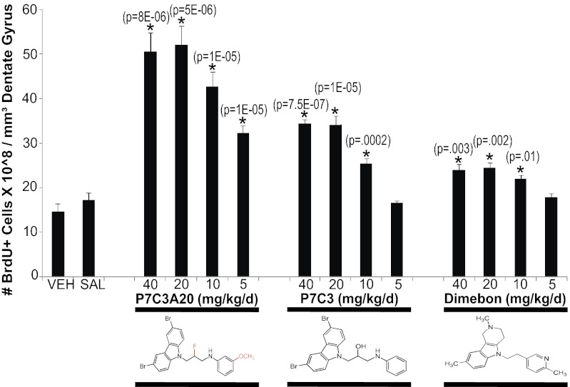
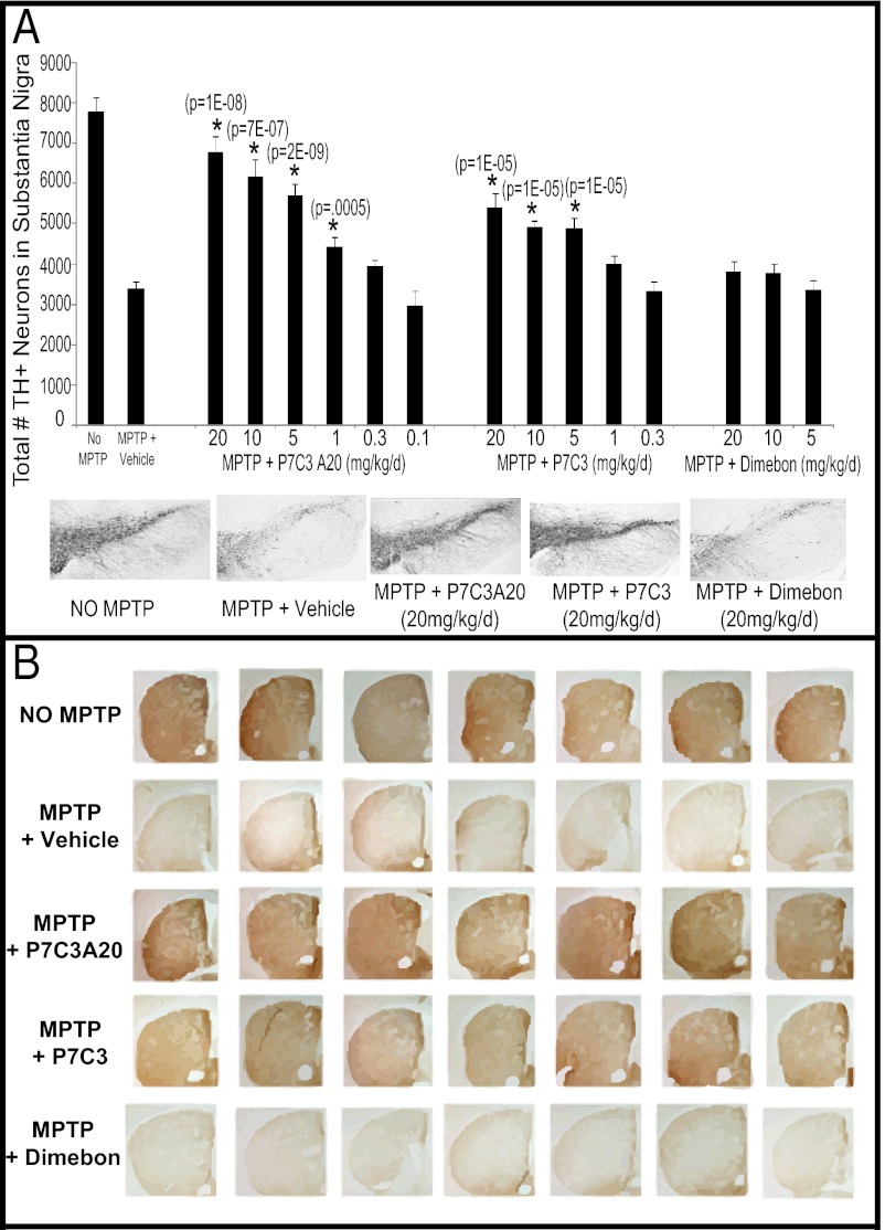
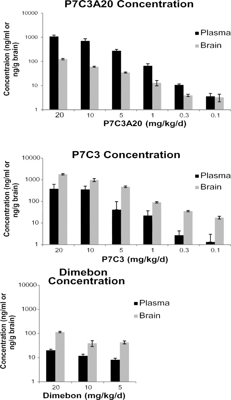
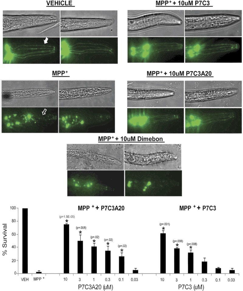
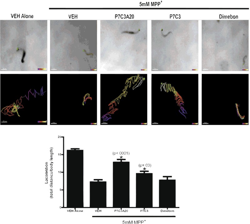
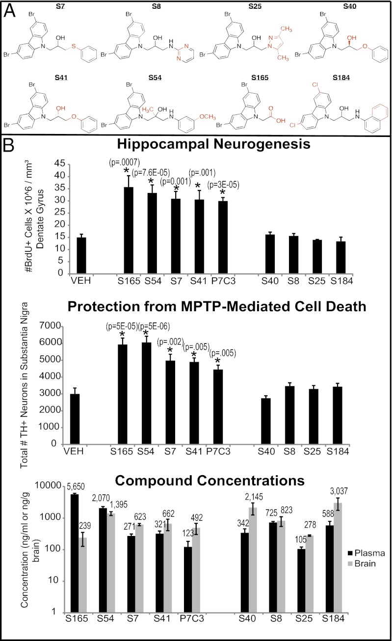
We previously reported the discovery of P7C3, an aminopropyl carbazole having proneurogenic and neuroprotective properties in newborn neural precursor cells of the dentate gyrus. Here, we provide evidence that P7C3 also protects mature neurons in brain regions outside of the hippocampus. P7C3 blocks 1-methyl-4-phenyl-1,2,3,6-tetrahydropyridine (MPTP)-mediated cell death of dopaminergic neurons in the substantia nigra of adult mice, a model of Parkinson disease (PD). Dose-response studies show that the P7C3 analog P7C3A20 blocks cell death with even greater potency and efficacy, which parallels the relative potency and efficacy of these agents in blocking apoptosis of newborn neural precursor cells of the dentate gyrus. P7C3 and P7C3A20 display similar relative effects in blocking 1-methyl-4-phenylpyridinium (MPP(+))-mediated death of dopaminergic neurons in Caenorhabditis elegans, as well as in preserving C. elegans mobility following MPP(+) exposure. Dimebon, an antihistaminergic drug that is weakly proneurogenic and neuroprotective in the dentate gyrus, confers no protection in either the mouse or the worm models of PD. We further demonstrate that the hippocampal proneurogenic efficacy of eight additional analogs of P7C3 correlates with their protective efficacy in MPTP-mediated neurotoxicity. In vivo screening of P7C3 analogs for proneurogenic efficacy in the hippocampus may thus provide a reliable means of predicting neuroprotective efficacy. We propose that the chemical scaffold represented by P7C3 and P7C3A20 provides a basis for optimizing and advancing pharmacologic agents for the treatment of patients with PD.


Conflict of interest statement

The authors declare no conflict of interest.

Figures

Fig. 1.
Fig. 1.
Neuroprotective efficacy of P7C3, P7C3A20, and Dimebon for newborn neurons in the adult hippocampus. Test compounds were evaluated by dose–response assay for their ability to block normal apoptotic cell death of newborn neural precursor cells in the adult dentate gyrus. P7C3A20 exhibits the greatest potency and ceiling of efficacy, and Dimebon the least. P7C3 is intermediate in both measures. Six animals were tested per group. Dosing is expressed as total milligrams per day, and compounds were administered intraperitoneally in divided doses twice daily. Data are expressed as mean ± SEM. Values for P7C3 and P7C3A20 were compared with those of vehicle (VEH), and values for Dimebon were compared with those of saline (SAL).
Fig. 2.
Fig. 2.
Neuroprotective efficacy of P7C3 and P7C3A20 from MPTP toxicity to SNc dopaminergic neurons. (A) Test compounds were evaluated by dose–response assay for their ability to block MPTP neurotoxicity in the SNc. P7C3A20 showed greater potency and CoE than P7C3, and Dimebon showed no protective efficacy. Fifteen animals were tested per group. The VEH group contained 30 animals: 15 animals that received the P7C3A20/P7C3 vehicle and 15 animals that received Dimebon vehicle (saline). These two control groups did not differ in number of surviving TH+ neurons and were thus combined. (Lower) Representative immunohistochemical pictures of TH staining in the SNc are shown. Dosing is expressed as total milligrams per day and was administered intraperitoneally in divided doses twice daily. Data are expressed as mean ± SEM. (B) Representative images of TH staining from the striata of individual animals demonstrate that 3 wk after a 5-d course of daily MPTP administration both P7C3 and P7C3A20 block the loss of dopaminergic axon terminals. P7C3A20 does so with greater effect, and Dimebon offers no protection. Striatal sections were obtained from the same mice used in Fig. 2, and compound treatment groups are from mice that received a dose of 20 mg⋅kg−1⋅d−1.
Fig. 3.
Fig. 3.
Brain and blood levels of P7C3, P7C3A20, and Dimebon 3 wk after MPTP administration. Relative neuroprotective activity within a test compound correlated with brain levels of that compound, and brain levels correlated with blood levels of the test compounds. Only about 1/10th the amount of P7C3A20 accumulated in the brain compared with P7C3. Brain accumulation of Dimebon was equivalent to that of P7C3. Data are expressed as mean ± SEM. Three animals were tested per group.
Fig. 4.
Fig. 4.
Neuroprotective efficacy of P7C3 and P7C3A20 for MPP+ toxicity to dopaminergic neurons in C. elegans. Worms were treated with 5 mM MPP+ for 40 h, with preincubation for 30 min with different concentrations of test compounds or vehicle. VEH animals not exposed to MPP+ exhibited normal GFP expression in dopaminergic neurons (solid arrow). By contrast, GFP expression was lost after 40 h of MPP+ exposure (open arrow). Both P7C3A20 and P7C3 dose dependently protected dopaminergic neurons from MPP+ toxicity, with P7C3A20 exhibiting greater potency and ceiling of efficacy. Twenty worms were analyzed per group, and each group analysis was performed in triplicate. Data are expressed as mean ± SEM.
Fig. 5.
Fig. 5.
Protective efficacy of P7C3 and P7C3A20 for MPP+-induced mobility deficits in C. elegans. (Top) Worms with the head identified by a green dot. (Middle) Path taken by each worm in 10 s, determined by tracking the green dot. Tracking is visualized as starting with blue color and progressing to white by the completion of 10 s. The green dot was used to determine locomotion, defined as the distance traveled by the head of the worm in 10 s divided by body length. (Scale bars, 70 μm.) Quantitative analysis of locomotion showed that untreated VEH controls had a value of 16.2 ± 0.49 (n = 30). When worms were treated with MPP+, locomotion was reduced more than 50% (7.2 ± 0.68; n = 31, P < 0.0001). A total of 10 μM P7C3A20 protected mobility to almost 80% of normal (12.8 ± 0.81; n = 34, *P < 0.01), and 10 μM P7C3 protected mobility almost 60% (mobility index = 9.6 ± 0.72; n = 28, *P < 0.05). A total of 10 μM Dimebon did not offer any protection (7.7 ± 1.0; n = 30). Experiments were performed in triplicate and data are expressed as mean ± SEM.
Fig. 6.
Fig. 6.
Efficacy of unique P7C3 analogs in the in vivo hippocampal neurogenesis assay correlates with activity in the in vivo MPTP-neuroprotection assay. (A) P7C3-S7 differs from P7C3 by replacing the aniline NH with sulfide linker. P7C3-S8 differs from P7C3 by replacing the aniline phenyl ring with a pyrimidine. P7C3-S25 differs from P7C3 by replacing the aniline moiety with a dimethyl pyrazole. P7C3-S40 and S41 differ from P7C3 by replacing the aniline NH with an oxygen linker, and they are R and S single enantiomers, respectively. P7C3-S54 differs from P7C3 mainly by the addition of a methyl group to the central carbon of the propyl linker and an OMe group on the aniline. P7C3-S165 differs from P7C3 by replacing the aniline and carbinol fragments with a carboxylic acid. P7C3-S184 differs from P7C3 by replacing the bromines on the carbazole with chlorines and replacing the aniline with a naphthyl amine. (B) Unique analogs of P7C3 were subjected to in vivo testing in the hippocampal neurogenesis (4 mice each) and MPTP-protection (10 mice each) assays. Results show that activity in these two assays correlates, such that the in vivo neurogenesis screen is useful for predicting neuroprotective efficacy of P7C3 analogs for dopaminergic neurons in the substantia nigra. LC/MS/MS assay of blood and brain levels of all compounds administered a single time to C57BL/6 mice at 10 mg/kg (i.p.) indicates that they cross the blood–brain barrier following i.p. administration.

Comment in

References

    1. Lees AJ, Hardy J, Revesz T. Parkinson’s disease. Lancet. 2009;373:2055–2066. - PubMed
    1. Pieper AA, et al. Discovery of a proneurogenic, neuroprotective chemical. Cell. 2010;142:39–51. - PMC - PubMed
    1. Pieper AA, et al. The neuronal PAS domain protein 3 transcription factor controls FGF-mediated adult hippocampal neurogenesis in mice. Proc Natl Acad Sci USA. 2005;102:14052–14057. - PMC - PubMed
    1. MacMillan KS, et al. Development of proneurogenic, neuroprotective small molecules. J Am Chem Soc. 2011;133:1428–1437. - PMC - PubMed
    1. McKnight SL, Pieper AA, Ready JM, De Brabander J. 2010. Proneurogenic compounds. US Patent 2010/020681.

Publication types

MeSH terms