

Comment
doi: 10.1073/pnas.1214903109.
Epub 2012 Oct 1.
Structure of the primed paramyxovirus fusion protein
Affiliations
- PMID: 23027936
- PMCID: PMC3478655
- DOI: 10.1073/pnas.1214903109
Item in Clipboard
Comment
Structure of the primed paramyxovirus fusion protein
Proc Natl Acad Sci U S A.
.
No abstract available
Conflict of interest statement
The authors declare no conflict of interest.
Figures
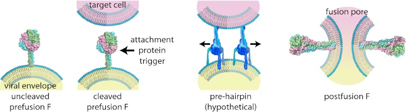
Cartoon of type I VFP-mediated viral entry, represented by example of the paramyxovirus F protein. Upon synthesis, uncleaved F assumes a metastable prefusion conformation (Left). Proteolytic maturation generates the fusion-ready prefusion form of the trimer (center, Left). Receptor binding by the associated attachment glycoprotein triggers major conformational changes of the primed F protein trimer. Refolding is considered to occur through a series of temporary conformations, including a hypothetical prehairpin intermediate (center, Right). Ultimately, the F protein assumes a thermodynamically highly stable postfusion conformation in which transmembrane domains and fusion peptides, and thus viral envelope and target membrane, are posited in close proximity (Right). Opening of a fusion pore enabling viral entry most likely requires concerted refolding of multiple F protein complexes. Structural renderings are based on original crystal structures [uncleaved prefusion F protein, Protein Data Bank (PDB) accession no. 2B9B; cleaved prefusion F protein from Welsh et al. (8); postfusion F protein, PDB accession no. 1ZTM) or hypothetical structural models (F protein prehairpin intermediate).
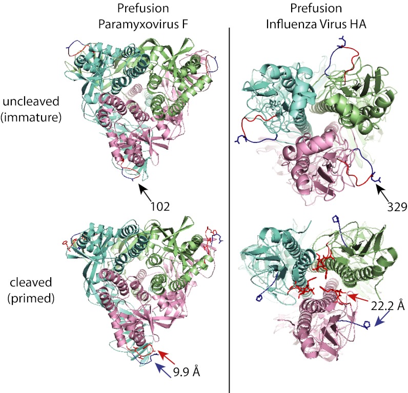
Ribbon representations of immature and mature prefusion paramyxovirus F protein [Left, PDB accession no. 2B9B and Welsh et al. (8)] and influenza virus HA (Right, PDB accession nos. 1HA0 and 2IBX) protein trimers, colored by monomer. Residues immediately upstream to the cleavage sites are colored dark blue, whereas residues immediately downstream representing the N-terminal sections of the fusion peptides are shown in red. Numbers and arrows (Upper) specify residue upstream to the cleavage sites in one of the monomers. Upon proteolytic maturation, only minor relocation of the newly generated N- and C-termini are observed in paramyxovirus F protein, whereas the N-terminal region of the influenza virus HA fusion peptide inserts deeply into the trimer interior (arrows, Lower). Numbers represent distance between chain termini after cleavage.
Comment on
-
Structure of the cleavage-activated prefusion form of the parainfluenza virus 5 fusion protein.Proc Natl Acad Sci U S A. 2012 Oct 9;109(41):16672-7. doi: 10.1073/pnas.1213802109. Epub 2012 Sep 10. Proc Natl Acad Sci U S A. 2012. PMID: 23012473 Free PMC article.
References
-
- Skehel JJ, Wiley DC. Coiled coils in both intracellular vesicle and viral membrane fusion. Cell. 1998;95:871–874. - PubMed
-
- Bizebard T, et al. Structure of influenza virus haemagglutinin complexed with a neutralizing antibody. Nature. 1995;376:92–94. - PubMed
-
- Bullough PA, Hughson FM, Skehel JJ, Wiley DC. Structure of influenza haemagglutinin at the pH of membrane fusion. Nature. 1994;371:37–43. - PubMed
Publication types
MeSH terms
Substances
Grants and funding
LinkOut - more resources
Full Text Sources
